0
views
0
recommends
+1 Recommend
0 collections
    0
    shares
      • Record: found
      • Abstract: found
      • Article: found
      Is Open Access

      Relationship between white matter alterations and contamination subgroup in obsessive compulsive disorder: A diffusion tensor imaging study

      research-article

      Read this article at

      Bookmark
          There is no author summary for this article yet. Authors can add summaries to their articles on ScienceOpen to make them more accessible to a non-specialist audience.

          Abstract

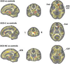
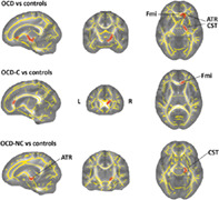
          Approximately 2%–3% of the world population suffers from obsessive–compulsive disorder (OCD). Several brain regions have been involved in the pathophysiology of OCD, but brain volumes in OCD may vary depending on specific OCD symptom dimensions. The study aims to explore how white matter structure changes in particular OCD symptom dimensions. Prior studies attempt to find the correlation between Y‐BOCS scores and OCD patients. However, in this study, we separated the contamination subgroup in OCD and compared directly to healthy control to find regions that exactly related to contamination symptoms. To evaluate structural alterations, diffusion tensor imaging was acquired from 30 OCD patients and 34 demographically matched healthy controls. Data were processed using tract‐based spatial statistics (TBSS) analysis. First, by comparing all OCD to healthy controls, significant fractional anisotropy (FA) decreased in the right anterior thalamic radiation, right corticospinal tract, and forceps minor observed. Then by comparing the contamination subgroup to healthy control, FA decreases in the forceps minor region. Consequently, forceps minor plays a central role in the pathophysiology of contamination behaviors. Finally, other subgroups were compared to healthy control and discovered that FA in the right corticospinal tract and right anterior thalamic radiation is reduced.

          Abstract

          In this study, we separated the contamination subgroup in obsessive–compulsive disorder and compared directly to healthy control to find regions that exactly related to contamination symptoms. Other subgroups were compared with healthy control and discovered that fractional anisotropy in the right corticospinal tract and right anterior thalamic radiation is reduced.

          Related collections

          Most cited references57

          • Record: found
          • Abstract: found
          • Article: not found

          Advances in functional and structural MR image analysis and implementation as FSL.

          The techniques available for the interrogation and analysis of neuroimaging data have a large influence in determining the flexibility, sensitivity, and scope of neuroimaging experiments. The development of such methodologies has allowed investigators to address scientific questions that could not previously be answered and, as such, has become an important research area in its own right. In this paper, we present a review of the research carried out by the Analysis Group at the Oxford Centre for Functional MRI of the Brain (FMRIB). This research has focussed on the development of new methodologies for the analysis of both structural and functional magnetic resonance imaging data. The majority of the research laid out in this paper has been implemented as freely available software tools within FMRIB's Software Library (FSL).
            Bookmark
            • Record: found
            • Abstract: found
            • Article: not found

            Tract-based spatial statistics: voxelwise analysis of multi-subject diffusion data.

            There has been much recent interest in using magnetic resonance diffusion imaging to provide information about anatomical connectivity in the brain, by measuring the anisotropic diffusion of water in white matter tracts. One of the measures most commonly derived from diffusion data is fractional anisotropy (FA), which quantifies how strongly directional the local tract structure is. Many imaging studies are starting to use FA images in voxelwise statistical analyses, in order to localise brain changes related to development, degeneration and disease. However, optimal analysis is compromised by the use of standard registration algorithms; there has not to date been a satisfactory solution to the question of how to align FA images from multiple subjects in a way that allows for valid conclusions to be drawn from the subsequent voxelwise analysis. Furthermore, the arbitrariness of the choice of spatial smoothing extent has not yet been resolved. In this paper, we present a new method that aims to solve these issues via (a) carefully tuned non-linear registration, followed by (b) projection onto an alignment-invariant tract representation (the "mean FA skeleton"). We refer to this new approach as Tract-Based Spatial Statistics (TBSS). TBSS aims to improve the sensitivity, objectivity and interpretability of analysis of multi-subject diffusion imaging studies. We describe TBSS in detail and present example TBSS results from several diffusion imaging studies.
              Bookmark
              • Record: found
              • Abstract: found
              • Article: not found

              MR diffusion tensor spectroscopy and imaging.

              This paper describes a new NMR imaging modality--MR diffusion tensor imaging. It consists of estimating an effective diffusion tensor, Deff, within a voxel, and then displaying useful quantities derived from it. We show how the phenomenon of anisotropic diffusion of water (or metabolites) in anisotropic tissues, measured noninvasively by these NMR methods, is exploited to determine fiber tract orientation and mean particle displacements. Once Deff is estimated from a series of NMR pulsed-gradient, spin-echo experiments, a tissue's three orthotropic axes can be determined. They coincide with the eigenvectors of Deff, while the effective diffusivities along these orthotropic directions are the eigenvalues of Deff. Diffusion ellipsoids, constructed in each voxel from Deff, depict both these orthotropic axes and the mean diffusion distances in these directions. Moreover, the three scalar invariants of Deff, which are independent of the tissue's orientation in the laboratory frame of reference, reveal useful information about molecular mobility reflective of local microstructure and anatomy. Inherently tensors (like Deff) describing transport processes in anisotropic media contain new information within a macroscopic voxel that scalars (such as the apparent diffusivity, proton density, T1, and T2) do not.
                Bookmark

                Author and article information

                Contributors
                karimi.m@iautmu.ac.ir , mkarimi.bme@gmail.com
                Journal
                Hum Brain Mapp
                Hum Brain Mapp
                10.1002/(ISSN)1097-0193
                HBM
                Human Brain Mapping
                John Wiley & Sons, Inc. (Hoboken, USA )
                1065-9471
                1097-0193
                27 March 2023
                1 June 2023
                : 44
                : 8 ( doiID: 10.1002/hbm.v44.8 )
                : 3302-3310
                Affiliations
                [ 1 ] Department of Biomedical Engineering, Faculty of Health, Tehran Medical Sciences Islamic Azad University Tehran Iran
                [ 2 ] School of Biotechnology and Biomolecular Sciences, Faculty of Science University of New South Wales (UNSW) Sydney, NSW Australia
                Author notes
                [*] [* ] Correspondence

                Mohammad Karimi Moridani, Department of Biomedical Engineering, Faculty of Health, Tehran Medical Sciences, Islamic Azad University, Tehran, Iran.

                Email: karimi.m@ 123456iautmu.ac.ir ; mkarimi.bme@ 123456gmail.com

                Author information
                https://orcid.org/0009-0003-9290-3601
                https://orcid.org/0000-0003-0793-3797
                https://orcid.org/0009-0004-0145-0537
                Article
                HBM26282
                10.1002/hbm.26282
                10171548
                36971658
                68f6ae6c-bbeb-4221-82c2-64fae30fbe74
                © 2023 The Authors. Human Brain Mapping published by Wiley Periodicals LLC.

                This is an open access article under the terms of the http://creativecommons.org/licenses/by/4.0/ License, which permits use, distribution and reproduction in any medium, provided the original work is properly cited.

                History
                : 27 January 2023
                : 25 June 2022
                : 06 March 2023
                Page count
                Figures: 4, Tables: 2, Pages: 9, Words: 8445
                Categories
                Research Article
                Research Articles
                Custom metadata
                2.0
                June 1, 2023
                Converter:WILEY_ML3GV2_TO_JATSPMC version:6.2.8 mode:remove_FC converted:10.05.2023

                Neurology
                contamination,diffusion tensor imaging,forceps minor,fractional anisotropy,obsessive–compulsive disorder,symptoms dimensions

                Comments

                Comment on this article