3
views
0
recommends
+1 Recommend
1 collections
    0
    shares
      • Record: found
      • Abstract: found
      • Article: found
      Is Open Access

      Development and external validation of a prediction model for the transition from mild to moderate or severe form of COVID-19

      research-article

      Read this article at

      Bookmark
          There is no author summary for this article yet. Authors can add summaries to their articles on ScienceOpen to make them more accessible to a non-specialist audience.

          Abstract

          Objectives

          COVID-19 pandemic seems to be under control. However, despite the vaccines, 5 to 10% of the patients with mild disease develop moderate to critical forms with potential lethal evolution. In addition to assess lung infection spread, chest CT helps to detect complications. Developing a prediction model to identify at-risk patients of worsening from mild COVID-19 combining simple clinical and biological parameters with qualitative or quantitative data using CT would be relevant to organizing optimal patient management.

          Methods

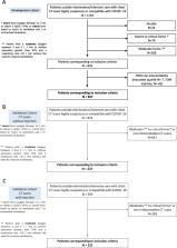
          Four French hospitals were used for model training and internal validation. External validation was conducted in two independent hospitals. We used easy-to-obtain clinical (age, gender, smoking, symptoms’ onset, cardiovascular comorbidities, diabetes, chronic respiratory diseases, immunosuppression) and biological parameters (lymphocytes, CRP) with qualitative or quantitative data (including radiomics) from the initial CT in mild COVID-19 patients.

          Results

          Qualitative CT scan with clinical and biological parameters can predict which patients with an initial mild presentation would develop a moderate to critical form of COVID-19, with a c-index of 0.70 (95% CI 0.63; 0.77). CT scan quantification improved the performance of the prediction up to 0.73 (95% CI 0.67; 0.79) and radiomics up to 0.77 (95% CI 0.71; 0.83). Results were similar in both validation cohorts, considering CT scans with or without injection.

          Conclusion

          Adding CT scan quantification or radiomics to simple clinical and biological parameters can better predict which patients with an initial mild COVID-19 would worsen than qualitative analyses alone. This tool could help to the fair use of healthcare resources and to screen patients for potential new drugs to prevent a pejorative evolution of COVID-19.

          Clinical Trial Registration

          NCT04481620.

          Clinical relevance statement

          CT scan quantification or radiomics analysis is superior to qualitative analysis, when used with simple clinical and biological parameters, to determine which patients with an initial mild presentation of COVID-19 would worsen to a moderate to critical form.

          Key Points

          • Qualitative CT scan analyses with simple clinical and biological parameters can predict which patients with an initial mild COVID-19 and respiratory symptoms would worsen with a c-index of 0.70.

          • Adding CT scan quantification improves the performance of the clinical prediction model to an AUC of 0.73.

          • Radiomics analyses slightly improve the performance of the model to a c-index of 0.77.

          Supplementary information

          The online version contains supplementary material available at 10.1007/s00330-023-09759-x.

          Related collections

          Most cited references33

          • Record: found
          • Abstract: found
          • Article: not found

          The REDCap consortium: Building an international community of software platform partners

          The Research Electronic Data Capture (REDCap) data management platform was developed in 2004 to address an institutional need at Vanderbilt University, then shared with a limited number of adopting sites beginning in 2006. Given bi-directional benefit in early sharing experiments, we created a broader consortium sharing and support model for any academic, non-profit, or government partner wishing to adopt the software. Our sharing framework and consortium-based support model have evolved over time along with the size of the consortium (currently more than 3200 REDCap partners across 128 countries). While the "REDCap Consortium" model represents only one example of how to build and disseminate a software platform, lessons learned from our approach may assist other research institutions seeking to build and disseminate innovative technologies.
            Bookmark
            • Record: found
            • Abstract: found
            • Article: not found

            Dexamethasone in Hospitalized Patients with Covid-19 — Preliminary Report

            Abstract Background Coronavirus disease 2019 (Covid-19) is associated with diffuse lung damage. Glucocorticoids may modulate inflammation-mediated lung injury and thereby reduce progression to respiratory failure and death. Methods In this controlled, open-label trial comparing a range of possible treatments in patients who were hospitalized with Covid-19, we randomly assigned patients to receive oral or intravenous dexamethasone (at a dose of 6 mg once daily) for up to 10 days or to receive usual care alone. The primary outcome was 28-day mortality. Here, we report the preliminary results of this comparison. Results A total of 2104 patients were assigned to receive dexamethasone and 4321 to receive usual care. Overall, 482 patients (22.9%) in the dexamethasone group and 1110 patients (25.7%) in the usual care group died within 28 days after randomization (age-adjusted rate ratio, 0.83; 95% confidence interval [CI], 0.75 to 0.93; P<0.001). The proportional and absolute between-group differences in mortality varied considerably according to the level of respiratory support that the patients were receiving at the time of randomization. In the dexamethasone group, the incidence of death was lower than that in the usual care group among patients receiving invasive mechanical ventilation (29.3% vs. 41.4%; rate ratio, 0.64; 95% CI, 0.51 to 0.81) and among those receiving oxygen without invasive mechanical ventilation (23.3% vs. 26.2%; rate ratio, 0.82; 95% CI, 0.72 to 0.94) but not among those who were receiving no respiratory support at randomization (17.8% vs. 14.0%; rate ratio, 1.19; 95% CI, 0.91 to 1.55). Conclusions In patients hospitalized with Covid-19, the use of dexamethasone resulted in lower 28-day mortality among those who were receiving either invasive mechanical ventilation or oxygen alone at randomization but not among those receiving no respiratory support. (Funded by the Medical Research Council and National Institute for Health Research and others; RECOVERY ClinicalTrials.gov number, NCT04381936; ISRCTN number, 50189673.)
              Bookmark
              • Record: found
              • Abstract: found
              • Article: not found

              Computational Radiomics System to Decode the Radiographic Phenotype

              Radiomics aims to quantify phenotypic characteristics on medical imaging through the use of automated algorithms. Radiomic artificial intelligence (AI) technology, either based on engineered hard-coded algorithms or deep learning methods, can be used to develop non-invasive imaging-based biomarkers. However, lack of standardized algorithm definitions and image processing severely hampers reproducibility and comparability of results. To address this issue, we developed PyRadiomics , a flexible open-source platform capable of extracting a large panel of engineered features from medical images. PyRadiomics is implemented in Python and can be used standalone or using 3D-Slicer. Here, we discuss the workflow and architecture of PyRadiomics and demonstrate its application in characterizing lung-lesions. Source code, documentation, and examples are publicly available at www.radiomics.io . With this platform, we aim to establish a reference standard for radiomic analyses, provide a tested and maintained resource, and to grow the community of radiomic developers addressing critical needs in cancer research.
                Bookmark

                Author and article information

                Contributors
                maeva.zysman@chu-bordeaux.fr
                Journal
                Eur Radiol
                Eur Radiol
                European Radiology
                Springer Berlin Heidelberg (Berlin/Heidelberg )
                0938-7994
                1432-1084
                5 July 2023
                5 July 2023
                2023
                : 33
                : 12
                : 9262-9274
                Affiliations
                [1 ]GRID grid.42399.35, ISNI 0000 0004 0593 7118, CHU Bordeaux, ; 33600 Pessac, France
                [2 ]GRID grid.503199.7, ISNI 0000 0004 0520 3579, Univ. Bordeaux, Centre de Recherche Cardio-Thoracique de Bordeaux, ; 33600 Bordeaux, France
                [3 ]GRID grid.457371.3, Centre de Recherche Cardio-Thoracique de Bordeaux (U1045), Centre d’Investigation Clinique, , INSERM, Bordeaux Population Health (U1219), ; (CIC-P 1401), 33600 Pessac, France
                [4 ]GRID grid.462496.b, ISNI 0000 0001 2302 4783, “Institut de Mathématiques de Bordeaux” (IMB), UMR5251, , CNRS, University of Bordeaux, ; 351 Cours Libération, 33400 Talence, France
                [5 ]GRID grid.457350.0, MONC Team & SISTM Team, , INRIA Bordeaux Sud-Ouest, ; 200 Av Vieille Tour, 33400 Talence, France
                [6 ]Pôle Des Spécialités Médicales/Département de Pneumologie, Université de Lorraine, Centre Hospitalier Régional Universitaire (CHRU) Nancy, ( https://ror.org/04vfs2w97) Service de Radiologie Et d’Imagerie, Nancy, France
                [7 ]Faculté de Médecine de Nancy, Université de Lorraine, Institut National de La Santé Et de La Recherche Médicale (INSERM) Unité Médicale de Recherche (UMR), ( https://ror.org/04vfs2w97) S 1116, Vandœuvre-Lès-Nancy, France
                [8 ]GRID grid.413745.0, ISNI 0000 0001 0507 738X, Department of Respiratory Diseases, , Arnaud de Villeneuve Hospital, Montpellier University Hospital, ; CEDEX 5, 34295 Montpellier, France
                [9 ]PhyMedExp, University of Montpellier, INSERM U1046, ( https://ror.org/051escj72) CEDEX 5, 34295 Montpellier, France
                [10 ]Pneumology Clinic, St Médard en Jalles, France
                [11 ]France Service de Radiologie Diagnostique Et Interventionnelle, Université Grenoble Alpes, CHU Grenoble-Alpes, ( https://ror.org/02rx3b187) Grenoble, France
                Article
                9759
                10.1007/s00330-023-09759-x
                10667132
                37405504
                6874f294-5864-47f9-862d-4a2d7473ad0f
                © The Author(s) 2023

                Open Access This article is licensed under a Creative Commons Attribution 4.0 International License, which permits use, sharing, adaptation, distribution and reproduction in any medium or format, as long as you give appropriate credit to the original author(s) and the source, provide a link to the Creative Commons licence, and indicate if changes were made. The images or other third party material in this article are included in the article's Creative Commons licence, unless indicated otherwise in a credit line to the material. If material is not included in the article's Creative Commons licence and your intended use is not permitted by statutory regulation or exceeds the permitted use, you will need to obtain permission directly from the copyright holder. To view a copy of this licence, visit http://creativecommons.org/licenses/by/4.0/.

                History
                : 1 December 2022
                : 22 March 2023
                : 4 April 2023
                Funding
                Funded by: phrci
                Categories
                Computed Tomography
                Custom metadata
                © European Society of Radiology 2023

                Radiology & Imaging
                covid-19,tomography, x-ray computed,clinical decision rules,artificial intelligence

                Comments

                Comment on this article