10
views
0
recommends
+1 Recommend
0 collections
    0
    shares
      • Record: found
      • Abstract: found
      • Article: found
      Is Open Access

      Tumor-infiltrating mast cells are associated with resistance to anti-PD-1 therapy

      research-article
      1 , , 1 , 1 , 1 , 1 , 1 , 1 , 1 , 2 , 1 , 1 , 1 , 1 , 1 , 1 , 1 , 1 , 3 , 4 , 4 , 5 , 5 , 3 , 1 , 1 , 1 , 1 , 1 , 1 , 1 , 1 , 1 , 1 , 1 , 1 , 1 , 1 , 1 , 1 , 1 , 1 , 6 , 1 , 1 , 1 , 1 , 1 , 1 , 7 , 7 , 8 , 9 , 10 , 1 , 12 , 8 , 11 , 1 ,
      Nature Communications
      Nature Publishing Group UK
      Melanoma, Tumour immunology, Adaptive immunity, Mast cells

      Read this article at

      Bookmark
          There is no author summary for this article yet. Authors can add summaries to their articles on ScienceOpen to make them more accessible to a non-specialist audience.

          Abstract

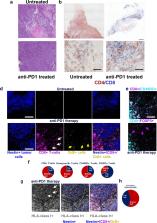
          Anti-PD-1 therapy is used as a front-line treatment for many cancers, but mechanistic insight into this therapy resistance is still lacking. Here we generate a humanized (Hu)-mouse melanoma model by injecting fetal liver-derived CD34 + cells and implanting autologous thymus in immune-deficient NOD- scid IL2Rγ null (NSG) mice. Reconstituted Hu-mice are challenged with HLA-matched melanomas and treated with anti-PD-1, which results in restricted tumor growth but not complete regression. Tumor RNA-seq, multiplexed imaging and immunohistology staining show high expression of chemokines, as well as recruitment of FOXP3 + Treg and mast cells, in selective tumor regions. Reduced HLA-class I expression and CD8 +/Granz B + T cells homeostasis are observed in tumor regions where FOXP3 + Treg and mast cells co-localize, with such features associated with resistance to anti-PD-1 treatment. Combining anti-PD-1 with sunitinib or imatinib results in the depletion of mast cells and complete regression of tumors. Our results thus implicate mast cell depletion for improving the efficacy of anti-PD-1 therapy.

          Abstract

          Immune checkpoint therapies (ICT) are promising for treating various cancers, but response rates vary. Here the authors show, in mouse models, that tumor-infiltrating mast cells colocalize with regulatory T cells, coincide with local reduction of MHC-I and CD8 T cells, and is associated with resistance to ICT, which can be reversed by c-kit inhibitor treatment.

          Related collections

          Most cited references46

          • Record: found
          • Abstract: found
          • Article: found
          Is Open Access

          Moderated estimation of fold change and dispersion for RNA-seq data with DESeq2

          In comparative high-throughput sequencing assays, a fundamental task is the analysis of count data, such as read counts per gene in RNA-seq, for evidence of systematic changes across experimental conditions. Small replicate numbers, discreteness, large dynamic range and the presence of outliers require a suitable statistical approach. We present DESeq2, a method for differential analysis of count data, using shrinkage estimation for dispersions and fold changes to improve stability and interpretability of estimates. This enables a more quantitative analysis focused on the strength rather than the mere presence of differential expression. The DESeq2 package is available at http://www.bioconductor.org/packages/release/bioc/html/DESeq2.html. Electronic supplementary material The online version of this article (doi:10.1186/s13059-014-0550-8) contains supplementary material, which is available to authorized users.
            Bookmark
            • Record: found
            • Abstract: found
            • Article: not found

            Primary, Adaptive, and Acquired Resistance to Cancer Immunotherapy.

            Cancer immunotherapy can induce long lasting responses in patients with metastatic cancers of a wide range of histologies. Broadening the clinical applicability of these treatments requires an improved understanding of the mechanisms limiting cancer immunotherapy. The interactions between the immune system and cancer cells are continuous, dynamic, and evolving from the initial establishment of a cancer cell to the development of metastatic disease, which is dependent on immune evasion. As the molecular mechanisms of resistance to immunotherapy are elucidated, actionable strategies to prevent or treat them may be derived to improve clinical outcomes for patients.
              Bookmark
              • Record: found
              • Abstract: found
              • Article: not found

              B cells and tertiary lymphoid structures promote immunotherapy response

              Treatment with immune checkpoint blockade (ICB) has revolutionized cancer therapy. Until now, predictive biomarkers1-10 and strategies to augment clinical response have largely focused on the T cell compartment. However, other immune subsets may also contribute to anti-tumour immunity11-15, although these have been less well-studied in ICB treatment16. A previously conducted neoadjuvant ICB trial in patients with melanoma showed via targeted expression profiling17 that B cell signatures were enriched in the tumours of patients who respond to treatment versus non-responding patients. To build on this, here we performed bulk RNA sequencing and found that B cell markers were the most differentially expressed genes in the tumours of responders versus non-responders. Our findings were corroborated using a computational method (MCP-counter18) to estimate the immune and stromal composition in this and two other ICB-treated cohorts (patients with melanoma and renal cell carcinoma). Histological evaluation highlighted the localization of B cells within tertiary lymphoid structures. We assessed the potential functional contributions of B cells via bulk and single-cell RNA sequencing, which demonstrate clonal expansion and unique functional states of B cells in responders. Mass cytometry showed that switched memory B cells were enriched in the tumours of responders. Together, these data provide insights into the potential role of B cells and tertiary lymphoid structures in the response to ICB treatment, with implications for the development of biomarkers and therapeutic targets.
                Bookmark

                Author and article information

                Contributors
                Shyam@wistar.org
                Herlynm@wistar.org
                Journal
                Nat Commun
                Nat Commun
                Nature Communications
                Nature Publishing Group UK (London )
                2041-1723
                12 January 2021
                12 January 2021
                2021
                : 12
                : 346
                Affiliations
                [1 ]GRID grid.251075.4, ISNI 0000 0001 1956 6678, The Wistar Institute, ; Philadelphia, PA USA
                [2 ]GRID grid.48336.3a, ISNI 0000 0004 1936 8075, Division of Cancer Epidemiology and Genetics, , National Cancer Institute, ; Bethesda, MD USA
                [3 ]GRID grid.25879.31, ISNI 0000 0004 1936 8972, Department of Pathology and Medicine, , University of Pennsylvania, ; Philadelphia, PA USA
                [4 ]GRID grid.25879.31, ISNI 0000 0004 1936 8972, Department of Medicine, Stem Cell and Xenograft Core, , University of Pennsylvania, ; Philadelphia, PA USA
                [5 ]GRID grid.25879.31, ISNI 0000 0004 1936 8972, Department of Genetics and Institute for Diabetes, Obesity and Metabolism, , Perelman School of Medicine, University of Pennsylvania, ; Philadelphia, PA USA
                [6 ]GRID grid.22937.3d, ISNI 0000 0000 9259 8492, Division of Immunology, Allergy and Infectious Diseases (DIAID), Department of Dermatology, , Medical University of Vienna, ; Vienna, Austria
                [7 ]GRID grid.5718.b, ISNI 0000 0001 2187 5445, University of Duisburg-Essen, ; Essen, Germany
                [8 ]GRID grid.240145.6, ISNI 0000 0001 2291 4776, Department of Surgical Oncology, , MD Anderson Cancer Center, ; Houston, TX USA
                [9 ]GRID grid.266102.1, ISNI 0000 0001 2297 6811, Department of Melanoma Medical Oncology, , University of California, ; San Francisco, CA USA
                [10 ]GRID grid.266102.1, ISNI 0000 0001 2297 6811, Department of Pathology and Dermatology, , University of California, ; San Francisco, CA USA
                [11 ]GRID grid.418152.b, AstraZeneca, ; Gaithersburg, MD USA
                [12 ]Present Address: GeneOne Life Science Inc., Fort Washington, PA USA
                Author information
                http://orcid.org/0000-0003-1553-4366
                http://orcid.org/0000-0002-5671-928X
                http://orcid.org/0000-0002-9949-798X
                http://orcid.org/0000-0002-7387-2219
                http://orcid.org/0000-0003-2206-9511
                http://orcid.org/0000-0002-2232-8512
                http://orcid.org/0000-0001-5799-6759
                http://orcid.org/0000-0001-9964-580X
                http://orcid.org/0000-0003-3729-2249
                http://orcid.org/0000-0002-0977-0912
                http://orcid.org/0000-0002-6807-2065
                http://orcid.org/0000-0003-3438-7576
                http://orcid.org/0000-0001-9913-6407
                Article
                20600
                10.1038/s41467-020-20600-7
                7804257
                33436641
                676ee912-f9ff-4ccc-a3e7-6be1aa2f75a8
                © The Author(s) 2021

                Open Access This article is licensed under a Creative Commons Attribution 4.0 International License, which permits use, sharing, adaptation, distribution and reproduction in any medium or format, as long as you give appropriate credit to the original author(s) and the source, provide a link to the Creative Commons license, and indicate if changes were made. The images or other third party material in this article are included in the article’s Creative Commons license, unless indicated otherwise in a credit line to the material. If material is not included in the article’s Creative Commons license and your intended use is not permitted by statutory regulation or exceeds the permitted use, you will need to obtain permission directly from the copyright holder. To view a copy of this license, visit http://creativecommons.org/licenses/by/4.0/.

                History
                : 16 April 2020
                : 10 December 2020
                Funding
                Funded by: FundRef https://doi.org/10.13039/100000002, U.S. Department of Health & Human Services | National Institutes of Health (NIH);
                Award ID: P50 CA174523, U54 CA224070
                Award ID: CA010815
                Award Recipient :
                Funded by: FundRef https://doi.org/10.13039/100000005, U.S. Department of Defense (United States Department of Defense);
                Award ID: PRCRP WX1XWH-16-1-0119 [CA150619], W81XWH-16-1-0120 and W81XWH-16-1-0121
                Award Recipient :
                Funded by: FundRef https://doi.org/10.13039/100005984, Dr. Miriam and Sheldon G. Adelson Medical Research Foundation (Dr. Miriam & Sheldon G. Adelson Medical Research Foundation);
                Funded by: U.S. Department of Health & Human Services | National Institutes of Health (NIH)
                Categories
                Article
                Custom metadata
                © The Author(s) 2021

                Uncategorized
                melanoma,tumour immunology,adaptive immunity,mast cells
                Uncategorized
                melanoma, tumour immunology, adaptive immunity, mast cells

                Comments

                Comment on this article