35
views
0
recommends
+1 Recommend
0 collections
    0
    shares
      • Record: found
      • Abstract: found
      • Article: found
      Is Open Access

      Alterations in adaptive immunity persist during long-duration spaceflight

      research-article

      Read this article at

      Bookmark
          There is no author summary for this article yet. Authors can add summaries to their articles on ScienceOpen to make them more accessible to a non-specialist audience.

          Abstract

          Background:

          It is currently unknown whether immune system alterations persist during long-duration spaceflight. In this study various adaptive immune parameters were assessed in astronauts at three intervals during 6-month spaceflight on board the International Space Station (ISS).

          AIMS:

          To assess phenotypic and functional immune system alterations in astronauts participating in 6-month orbital spaceflight.

          Methods:

          Blood was collected before, during, and after flight from 23 astronauts participating in 6-month ISS expeditions. In-flight samples were returned to Earth within 48 h of collection for immediate analysis. Assays included peripheral leukocyte distribution, T-cell function, virus-specific immunity, and mitogen-stimulated cytokine production profiles.

          Results:

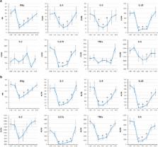
          Redistribution of leukocyte subsets occurred during flight, including an elevated white blood cell (WBC) count and alterations in CD8 + T-cell maturation. A reduction in general T-cell function (both CD4 + and CD8 +) persisted for the duration of the 6-month spaceflights, with differential responses between mitogens suggesting an activation threshold shift. The percentage of CD4 + T cells capable of producing IL-2 was depressed after landing. Significant reductions in mitogen-stimulated production of IFNγ, IL-10, IL-5, TNFα, and IL-6 persisted during spaceflight. Following lipopolysaccharide (LPS) stimulation, production of IL-10 was reduced, whereas IL-8 production was increased during flight.

          Conclusions:

          The data indicated that immune alterations persist during long-duration spaceflight. This phenomenon, in the absence of appropriate countermeasures, has the potential to increase specific clinical risks for crewmembers during exploration-class deep space missions.

          Related collections

          Most cited references40

          • Record: found
          • Abstract: found
          • Article: not found

          Immune system dysregulation following short- vs long-duration spaceflight.

          Immune system dysregulation has been demonstrated to occur during and immediately following spaceflight. If found to persist during lengthy flights, this phenomenon could be a serious health risk to crewmembers participating in lunar or Mars missions. A comprehensive postflight immune assessment was performed on 17 short-duration Space Shuttle crewmembers and 8 long-duration International Space Station (ISS) crewmembers. Testing consisted of peripheral leukocyte subset analysis, early T cell activation potential, and intracellular/secreted cytokine profiles. For Shuttle crewmembers, the distribution of the peripheral leukocyte subsets was found to be altered postflight. Early T cell activation was elevated postflight; however, the percentage of T cell subsets capable of being stimulated to produce IL-2 and IFN gamma was decreased. The ratio of secreted IFN gamma:IL-10 following T cell stimulation declined after landing, indicating a Th2 shift. For the ISS crewmembers, some alterations in peripheral leukocyte distribution were also detected after landing. In contrast to Shuttle crewmembers, the ISS crewmembers demonstrated a statistically significant reduction in early T cell activation potential immediately postflight. The percentage of T cells capable of producing IL-2 was reduced, but IFN gamma percentages were unchanged. A reduction in the secreted IFN gamma:IL-10 ratio (Th2 shift) was also observed postflight in the ISS crewmembers. These data indicate that consistent peripheral phenotype changes and altered cytokine production profiles occur following spaceflight of both short and long duration; however, functional immune dysregulation may vary related to mission duration. In addition, a detectable Th2 cytokine shift appears to be associated with spaceflight.
            Bookmark
            • Record: found
            • Abstract: found
            • Article: not found

            Stress-induced subclinical reactivation of varicella zoster virus in astronauts.

            Varicella zoster virus (VZV) becomes latent in human ganglia after primary infection. VZV reactivation occurs primarily in elderly individuals, organ transplant recipients, and patients with cancer and AIDS, correlating with a specific decline in cell-mediated immunity to the virus. VZV can also reactivate after surgical stress. The unexpected occurrence of thoracic zoster 2 days before space flight in a 47-year-old healthy astronaut from a pool of 81 physically fit astronauts prompted our search for VZV reactivation during times of stress to determine whether VZV can also reactivate after non-surgical stress. We examined total DNA extracted from 312 saliva samples of eight astronauts before, during, and after space flight for VZV DNA by polymerase chain reaction: 112 samples were obtained 234-265 days before flight, 84 samples on days 2 through 13 of space flight, and 116 samples on days 1 through 15 after flight. Before space flight, only one of the 112 saliva samples from a single astronaut was positive for VZV DNA. In contrast, during and after space flight, 61 of 200 (30%) saliva samples were positive in all eight astronauts. No VZV DNA was detected in any of 88 saliva samples from 10 healthy control subjects. These results indicate that VZV can reactivate subclinically in healthy individuals after non-surgical stress. Copyright 2004 Wiley-Liss, Inc.
              Bookmark
              • Record: found
              • Abstract: found
              • Article: not found

              T-cell triggering thresholds are modulated by the number of antigen within individual T-cell receptor clusters.

              T cells react to extremely small numbers of activating agonist peptides. Spatial organization of T-cell receptors (TCR) and their peptide-major histocompatibility complex (pMHC) ligands into microclusters is correlated with T-cell activation. Here we have designed an experimental strategy that enables control over the number of agonist peptides per TCR cluster, without altering the total number engaged by the cell. Supported membranes, partitioned with grids of barriers to lateral mobility, provide an effective way of limiting the total number of pMHC ligands that may be assembled within a single TCR cluster. Observations directly reveal that restriction of pMHC content within individual TCR clusters can decrease T-cell sensitivity for triggering initial calcium flux at fixed total pMHC density. Further analysis suggests that triggering thresholds are determined by the number of activating ligands available to individual TCR clusters, not by the total number encountered by the cell. Results from a series of experiments in which the overall agonist density and the maximum number of agonist per TCR cluster are independently varied in primary T cells indicate that the most probable minimal triggering unit for calcium signaling is at least four pMHC in a single cluster for this system. This threshold is unchanged by inclusion of coagonist pMHC, but costimulation of CD28 by CD80 can modulate the threshold lower.
                Bookmark

                Author and article information

                Journal
                NPJ Microgravity
                NPJ Microgravity
                NPJ Microgravity
                Nature Publishing Group
                2373-8065
                03 September 2015
                2015
                : 1
                : 15013
                Affiliations
                [1 ] Biomedical Research and Environmental Sciences Division, NASA Johnson Space Center , Houston, TX, USA
                [2 ] Microgen Laboratories , La Marque, TX, USA
                [3 ] Biomedical Research and Environmental Sciences Division, Enterprise Advisory Services, Inc. , Houston, TX, USA
                [4 ] Biomedical Research and Environmental Sciences Division, JES Tech , Houston, TX, USA
                [5 ] Space and Clinical Operations Division , Houston, TX, USA
                Author notes

                BC: study operational support, flow cytometry assays, manuscript preparation. RPS: virus-specific immunity assays. SM: Virus reactivation assays. HQ: Study operational support, flow cytometry assays. DP: virus reactivation assays. CS: principal investigator.

                Article
                npjmgrav201513
                10.1038/npjmgrav.2015.13
                5515498
                28725716
                675f10d7-5ea0-4981-a1e7-ba9497e4f388
                Copyright © 2015 Macmillan Publishers Limited

                This work is licensed under a Creative Commons Attribution-NonCommercial-NoDerivatives 4.0 International License. The images or other third party material in this article are included in the article’s Creative Commons license, unless indicated otherwise in the credit line; if the material is not included under the Creative Commons license, users will need to obtain permission from the license holder to reproduce the material. To view a copy of this license, visit http://creativecommons.org/licenses/by-nc-nd/4.0/

                History
                : 28 January 2015
                : 26 June 2015
                : 13 July 2015
                Categories
                Article

                Comments

                Comment on this article