37
views
0
recommends
+1 Recommend
0 collections
    0
    shares
      • Record: found
      • Abstract: found
      • Article: found
      Is Open Access

      Sound exposure dynamically induces dopamine synthesis in cholinergic LOC efferents for feedback to auditory nerve fibers

      research-article

      Read this article at

      Bookmark
          There is no author summary for this article yet. Authors can add summaries to their articles on ScienceOpen to make them more accessible to a non-specialist audience.

          Abstract

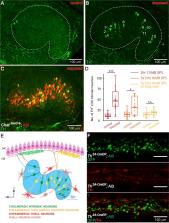
          Lateral olivocochlear (LOC) efferent neurons modulate auditory nerve fiber (ANF) activity using a large repertoire of neurotransmitters, including dopamine (DA) and acetylcholine (ACh). Little is known about how individual neurotransmitter systems are differentially utilized in response to the ever-changing acoustic environment. Here we present quantitative evidence in rodents that the dopaminergic LOC input to ANFs is dynamically regulated according to the animal’s recent acoustic experience. Sound exposure upregulates tyrosine hydroxylase, an enzyme responsible for dopamine synthesis, in cholinergic LOC intrinsic neurons, suggesting that individual LOC neurons might at times co-release ACh and DA. We further demonstrate that dopamine down-regulates ANF firing rates by reducing both the hair cell release rate and the size of synaptic events. Collectively, our results suggest that LOC intrinsic neurons can undergo on-demand neurotransmitter re-specification to re-calibrate ANF activity, adjust the gain at hair cell/ANF synapses, and possibly to protect these synapses from noise damage.

          eLife digest

          Every day, we hear sounds that might be alarming, distracting, intriguing or calming – or simply just too loud. Our hearing system responds to these acoustic changes by fine-tuning sounds before they enter the brain. For example, if a noise is too loud, the volume can be turned down by dampening the signals nerve fibers in the ear send to the brain. This is thought to reduce the damage loud sounds can cause to the sensory organ inside the ear.

          A set of nerve cells located at the base of the brain called the lateral olivocochlear (LOC) neurons coordinate this adjustment to different volumes and sounds. When these neurons receive information on external sounds, they signal back to the hearing organs and adjust the activity of auditory nerve fibers that communicate this information to the brain. LOC neurons use a diverse range of molecules to modify the activity of auditory nerve fibers, including the ‘feel-good’ neurotransmitter dopamine. But it is unclear what role dopamine plays in this auditory feedback loop.

          To find out, Wu et al. studied the hearing system of mice that had been exposed to different levels of sound. This involved imaging LOC neurons stained with a marker for dopamine and measuring the activity of nerve fibers in the inner ear. The experiments showed that LOC neurons in mice that had recently been exposed to sound were covered in an enzyme that is essential for making dopamine. The louder the sound, the more of this enzyme was present, suggesting that the amount of dopamine released depends on the volume of the sound.

          LOC neurons release another neurotransmitter called acetylcholine, which stimulates activity in auditory nerve fibers. Wu et al. found that dopamine and acetylcholine are released from the same group of LOC neurons. However, dopamine had the opposite effect to acetylcholine and reduced nerve activity. These findings suggest that by controlling the mixture of neurotransmitters released, LOC neurons are able to fine-tune the activity of auditory nerve fibers in response to acoustic changes.

          This work provides a new insight into how our hearing system is able to perceive and relay changes in the sound environment. A better understanding of this auditory feedback loop could influence the design of implant devices for people with impaired hearing.

          Related collections

          Most cited references77

          • Record: found
          • Abstract: found
          • Article: not found

          Melanocortin-4 receptors expressed by cholinergic neurons regulate energy balance and glucose homeostasis.

          Melanocortin-4 receptor (MC4R) mutations cause dysregulation of energy balance and hyperinsulinemia. We have used mouse models to study the physiological roles of extrahypothalamic MC4Rs. Re-expression of MC4Rs in cholinergic neurons (ChAT-Cre, loxTB MC4R mice) modestly reduced body weight gain without altering food intake and was sufficient to normalize energy expenditure and attenuate hyperglycemia and hyperinsulinemia. In contrast, restoration of MC4R expression in brainstem neurons including those in the dorsal motor nucleus of the vagus (Phox2b-Cre, loxTB MC4R mice) was sufficient to attenuate hyperinsulinemia, while the hyperglycemia and energy balance were not normalized. Additionally, hepatic insulin action and insulin-mediated suppression of hepatic glucose production were improved in ChAT-Cre, loxTB MC4R mice. These findings suggest that MC4Rs expressed by cholinergic neurons regulate energy expenditure and hepatic glucose production. Our results also provide further evidence of the dissociation in pathways mediating the effects of melanocortins on energy balance and glucose homeostasis. Copyright © 2011 Elsevier Inc. All rights reserved.
            Bookmark
            • Record: found
            • Abstract: found
            • Article: not found

            Transmitter release at the hair cell ribbon synapse.

            Neurotransmitters are released continuously at ribbon synapses in the retina and cochlea. Notably, a single ribbon synapse of inner hair cells provides the entire input to each cochlear afferent fiber. We investigated hair cell transmitter release in the postnatal rat cochlea by recording excitatory postsynaptic currents (EPSCs) from afferent boutons directly abutting the ribbon synapse. EPSCs were carried by rapidly gating AMPA receptors. EPSCs were clustered in time, indicating the possibility of coordinate release. Amplitude distributions of spontaneous EPSCs were highly skewed, peaking at 0.4 nS and ranging up to 20 times larger. Hair cell depolarization increased EPSC frequency up to 150 Hz without altering the amplitude distribution. We propose that the ribbon synapse operates by multivesicular release, possibly to achieve high-frequency transmission.
              Bookmark
              • Record: found
              • Abstract: found
              • Article: not found

              A physiological place-frequency map of the cochlea in the CBA/J mouse.

              Genetically manipulated mice have gained a prominent role in in vivo research on development and function of the auditory system. A prerequisite for the interpretation of normal and abnormal structural and functional features of the inner ear is the exact knowledge of the cochlear place-frequency map. Using a stereotaxic approach to the projection site of the auditory nerve fibers in the cochlear nucleus, we succeeded in labelling physiologically characterized auditory nerve afferents and determined their peripheral innervation site in the cochlea. From the neuronal characteristic frequency (CF) and the innervation site in the organ of Corti a place-frequency map was established for characteristic frequencies between 7.2 and 61.8 kHz, corresponding to locations between 90% and 10% basilar membrane length (base = 0%, apex = 100%, mean length measured under the inner hair cells 5.13 mm). The relation between normalized distance from the base (d) and frequency (kHz) can be described by a simple logarithmic function: d(%) = 156.5-82.5 x log(f), with a slope of 1.25 mm/octave of frequency. The present map, recorded under physiological conditions, differs from earlier maps determined with different methods. The simple logarithmic place-frequency relation found in the mouse indicates that mice are acoustic generalists rather than specialists.
                Bookmark

                Author and article information

                Contributors
                Role: Senior Editor
                Role: Reviewing Editor
                Journal
                eLife
                Elife
                eLife
                eLife
                eLife Sciences Publications, Ltd
                2050-084X
                24 January 2020
                2020
                : 9
                : e52419
                Affiliations
                [1 ]deptDepartment of Neuroscience The Johns Hopkins University School of Medicine BaltimoreUnited States
                [2 ]deptThe Center for Sensory Biology The Johns Hopkins University School of Medicine BaltimoreUnited States
                [3 ]deptThe Center for Hearing and Balance The Johns Hopkins University School of Medicine BaltimoreUnited States
                [4 ]College of Pharmacy and Natural Medicine Research Institute, Mokpo National University Muan-gunRepublic of Korea
                [5 ]deptDepartment of Otolaryngology-Head and Neck Surgery The Johns Hopkins University School of Medicine BaltimoreUnited States
                University of Oxford United Kingdom
                University of Maryland United States
                University of Maryland United States
                Harvard Medical School United States
                Rochester United States
                Author notes
                [†]

                Department of Neurobiology, Harvard Medical School, Boston, United States.

                Author information
                https://orcid.org/0000-0001-9625-1661
                http://orcid.org/0000-0003-4184-7374
                https://orcid.org/0000-0003-3135-658X
                Article
                52419
                10.7554/eLife.52419
                7043886
                31975688
                67011562-bb6a-4a51-9fcf-8a66b3700019
                © 2020, Wu et al

                This article is distributed under the terms of the Creative Commons Attribution License, which permits unrestricted use and redistribution provided that the original author and source are credited.

                History
                : 03 October 2019
                : 23 January 2020
                Funding
                Funded by: FundRef http://dx.doi.org/10.13039/100000055, National Institute on Deafness and Other Communication Disorders;
                Award ID: R01DC006476
                Award Recipient :
                Funded by: FundRef http://dx.doi.org/10.13039/100000055, National Institute on Deafness and Other Communication Disorders;
                Award ID: R01DC012957
                Award Recipient :
                Funded by: FundRef http://dx.doi.org/10.13039/100000055, National Institute on Deafness and Other Communication Disorders;
                Award ID: R01DC017620
                Award Recipient :
                Funded by: FundRef http://dx.doi.org/10.13039/100000055, National Institute on Deafness and Other Communication Disorders;
                Award ID: R01DC016641
                Award Recipient :
                Funded by: David M. Rubenstein Fund for Hearing Research;
                Award Recipient :
                Funded by: FundRef http://dx.doi.org/10.13039/100008180, Capita Foundation;
                Award Recipient :
                Funded by: FundRef http://dx.doi.org/10.13039/501100003710, Korea Health Industry Development Institute;
                Award ID: Korea Health Technology R&D Project HI17C0952
                Award Recipient :
                The funders had no role in study design, data collection and interpretation, or the decision to submit the work for publication.
                Categories
                Research Article
                Neuroscience
                Custom metadata
                Based on the animal’s recent history of sound exposure, cholinergic auditory brainstem neurons dynamically regulate dopamine synthesis for inhibitory feedback to the inner ear.

                Life sciences
                auditory nerve fiber,spiral ganglion neuron,olivocochlear efferents,dopamine,acetylcholine,noise damage,mouse,rat

                Comments

                Comment on this article