9
views
0
recommends
+1 Recommend
0 collections
    0
    shares
      • Record: found
      • Abstract: found
      • Article: found
      Is Open Access

      Interleukin-36 in Infectious and Inflammatory Skin Diseases

      review-article
      , *
      Frontiers in Immunology
      Frontiers Media S.A.
      IL-36, IL-36γ, inflammation, infection, skin, dermatosis, psoriasis

      Read this article at

      Bookmark
          There is no author summary for this article yet. Authors can add summaries to their articles on ScienceOpen to make them more accessible to a non-specialist audience.

          Abstract

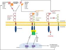
          Interleukin-36 (IL-36) comprises to a cytokine family consisting of four isoforms IL-36α, IL-36β, IL-36γ, and IL-36 receptor antagonist (IL-36 Ra). These IL-36 cytokines, in turn, belong to the IL-1 superfamily. The IL-36 receptor (IL-1R6) is functional as a heterodimer formed of IL-1R6 and IL-1 receptor accessory protein (IL-1RAcP). IL-36α, IL-36β, and IL-36γ are regarded as pro-inflammatory ligands and IL-36 Ra as well as IL-38 as anti-inflammatory ligands of IL-1R6. IL-36 cytokines are mainly expressed on the barrier sites of the body e.g., bronchial, intestinal, and dermal epithelium. One of their most important biological functions is the bridging of innate and adaptive immune responses. A disturbed balance between pro-inflammatory and anti-inflammatory branches easily leads to inflammation of the corresponding tissue. The most prominent example for an altered IL-36 expression is the spectrum of psoriasis. In addition to inflammatory dermatoses, IL-36 also seems to play a role in infectious dermatoses. Microbial triggers, especially Staphylococcus aureus infection, increase the production of pro-inflammatory IL-36 cytokines and initiate/promote the inflammation of skin lesions. Due to the discovery of IL-36 as an important immune mediator, it has already been possible to develop important diagnostic tools for dermatitis. Not only in the field of inflammatory skin diseases, but also in pulmonary and intestinal inflammation, there is evidence that IL-36 cytokines might have diagnostic and/or therapeutic relevance.

          Related collections

          Most cited references99

          • Record: found
          • Abstract: found
          • Article: not found

          Methicillin-resistant S. aureus infections among patients in the emergency department.

          Methicillin-resistant Staphylococcus aureus (MRSA) is increasingly recognized in infections among persons in the community without established risk factors for MRSA. We enrolled adult patients with acute, purulent skin and soft-tissue infections presenting to 11 university-affiliated emergency departments during the month of August 2004. Cultures were obtained, and clinical information was collected. Available S. aureus isolates were characterized by antimicrobial-susceptibility testing, pulsed-field gel electrophoresis, and detection of toxin genes. On MRSA isolates, we performed typing of the staphylococcal cassette chromosome mec (SCCmec), the genetic element that carries the mecA gene encoding methicillin resistance. S. aureus was isolated from 320 of 422 patients with skin and soft-tissue infections (76 percent). The prevalence of MRSA was 59 percent overall and ranged from 15 to 74 percent. Pulsed-field type USA300 isolates accounted for 97 percent of MRSA isolates; 74 percent of these were a single strain (USA300-0114). SCCmec type IV and the Panton-Valentine leukocidin toxin gene were detected in 98 percent of MRSA isolates. Other toxin genes were detected rarely. Among the MRSA isolates, 95 percent were susceptible to clindamycin, 6 percent to erythromycin, 60 percent to fluoroquinolones, 100 percent to rifampin and trimethoprim-sulfamethoxazole, and 92 percent to tetracycline. Antibiotic therapy was not concordant with the results of susceptibility testing in 100 of 175 patients with MRSA infection who received antibiotics (57 percent). Among methicillin-susceptible S. aureus isolates, 31 percent were USA300 and 42 percent contained pvl genes. MRSA is the most common identifiable cause of skin and soft-tissue infections among patients presenting to emergency departments in 11 U.S. cities. When antimicrobial therapy is indicated for the treatment of skin and soft-tissue infections, clinicians should consider obtaining cultures and modifying empirical therapy to provide MRSA coverage. Copyright 2006 Massachusetts Medical Society.
            Bookmark
            • Record: found
            • Abstract: found
            • Article: not found

            Interleukin-36-receptor antagonist deficiency and generalized pustular psoriasis.

            Generalized pustular psoriasis is a life-threatening disease of unknown cause. It is characterized by sudden, repeated episodes of high-grade fever, generalized rash, and disseminated pustules, with hyperleukocytosis and elevated serum levels of C-reactive protein, which may be associated with plaque-type psoriasis. We performed homozygosity mapping and direct sequencing in nine Tunisian multiplex families with autosomal recessive generalized pustular psoriasis. We assessed the effect of mutations on protein expression and conformation, stability, and function. We identified significant linkage to an interval of 1.2 megabases on chromosome 2q13-q14.1 and a homozygous missense mutation in IL36RN, encoding an interleukin-36-receptor antagonist (interleukin-36Ra), an antiinflammatory cytokine. This mutation predicts the substitution of a proline residue for leucine at amino acid position 27 (L27P). Homology-based structural modeling of human interleukin-36Ra suggests that the proline at position 27 affects both the stability of interleukin-36Ra and its interaction with its receptor, interleukin-1 receptor-like 2 (interleukin-1 receptor-related protein 2). Biochemical analyses showed that the L27P variant was poorly expressed and less potent than the nonvariant interleukin-36Ra in inhibiting a cytokine-induced response in an interleukin-8 reporter assay, leading to enhanced production of inflammatory cytokines (interleukin-8 in particular) by keratinocytes from the patients. Aberrant interleukin-36Ra structure and function lead to unregulated secretion of inflammatory cytokines and generalized pustular psoriasis. (Funded by Agence Nationale de la Recherche and Société Française de Dermatologie.).
              Bookmark
              • Record: found
              • Abstract: found
              • Article: not found

              The interleukin (IL)-1 cytokine family--Balance between agonists and antagonists in inflammatory diseases.

              The interleukin (IL)-1 family of cytokines comprises 11 members, including 7 pro-inflammatory agonists (IL-1α, IL-1β, IL-18, IL-33, IL-36α, IL-36β, IL-36γ) and 4 defined or putative antagonists (IL-1R antagonist (IL-1Ra), IL-36Ra, IL-37, and IL-38) exerting anti-inflammatory activities. Except for IL-1Ra, IL-1 cytokines do not possess a leader sequence and are secreted via an unconventional pathway. In addition, IL-1β and IL-18 are produced as biologically inert pro-peptides that require cleavage by caspase-1 in their N-terminal region to generate active proteins. N-terminal processing is also required for full activity of IL-36 cytokines. The IL-1 receptor (IL-1R) family comprises 10 members and includes cytokine-specific receptors, co-receptors and inhibitory receptors. The signaling IL-1Rs share a common structure with three extracellular immunoglobulin (Ig) domains and an intracellular Toll-like/IL-1R (TIR) domain. IL-1 cytokines bind to their specific receptor, which leads to the recruitment of a co-receptor and intracellular signaling. IL-1 cytokines induce potent inflammatory responses and their activity is tightly controlled at the level of production, protein processing and maturation, receptor binding and post-receptor signaling by naturally occurring inhibitors. Some of these inhibitors are IL-1 family antagonists, while others are IL-1R family members acting as membrane-bound or soluble decoy receptors. An imbalance between agonist and antagonist levels can lead to exaggerated inflammatory responses. Several genetic modifications or mutations associated with dysregulated IL-1 activity and autoinflammatory disorders were identified in mouse models and in patients. These findings paved the road to the successful use of IL-1 inhibitors in diseases that were previously considered as untreatable.
                Bookmark

                Author and article information

                Contributors
                Journal
                Front Immunol
                Front Immunol
                Front. Immunol.
                Frontiers in Immunology
                Frontiers Media S.A.
                1664-3224
                24 May 2019
                2019
                : 10
                : 1162
                Affiliations
                Department of Dermatology and Allergy, University Hospital of Bonn , Bonn, Germany
                Author notes

                Edited by: Ralf J. Ludwig, Universität zu Lübeck, Germany

                Reviewed by: Heiko Mühl, Goethe-Universität Frankfurt am Main, Germany; Unni Samavedam, University of Cincinnati, United States

                *Correspondence: Joerg Wenzel joerg.wenzel@ 123456ukbonn.de

                This article was submitted to Autoimmune and Autoinflammatory Disorders, a section of the journal Frontiers in Immunology

                Article
                10.3389/fimmu.2019.01162
                6545975
                31191535
                66733c56-0acc-41cc-a842-4fc8062a9723
                Copyright © 2019 Buhl and Wenzel.

                This is an open-access article distributed under the terms of the Creative Commons Attribution License (CC BY). The use, distribution or reproduction in other forums is permitted, provided the original author(s) and the copyright owner(s) are credited and that the original publication in this journal is cited, in accordance with accepted academic practice. No use, distribution or reproduction is permitted which does not comply with these terms.

                History
                : 03 April 2019
                : 08 May 2019
                Page count
                Figures: 3, Tables: 1, Equations: 0, References: 102, Pages: 11, Words: 8597
                Categories
                Immunology
                Review

                Immunology
                il-36,il-36γ,inflammation,infection,skin,dermatosis,psoriasis
                Immunology
                il-36, il-36γ, inflammation, infection, skin, dermatosis, psoriasis

                Comments

                Comment on this article