1
views
0
recommends
+1 Recommend
0 collections
    0
    shares
      • Record: found
      • Abstract: found
      • Article: found
      Is Open Access

      Artificial Intelligence for Hip Fracture Detection and Outcome Prediction : A Systematic Review and Meta-analysis

      research-article
      , MBChB 1 , 2 , 3 , , , MD 1 , , MD 2 , 4 , , MD, PhD 1 , 5 , , PhD 3 , , MD, PhD 1 , 5
      JAMA Network Open
      American Medical Association

      Read this article at

      Bookmark
          There is no author summary for this article yet. Authors can add summaries to their articles on ScienceOpen to make them more accessible to a non-specialist audience.

          Key Points

          Question

          For patients with hip fractures, how well do current artificial intelligence algorithms perform at diagnosing fractures and predicting postoperative outcomes?

          Findings

          This systematic review and meta-analysis of 39 studies identified similar error rates of hip fracture diagnosis between artificial intelligence models and expert clinicians. There was minimal advantage of machine learning models over traditional regression techniques for postoperative outcome prediction.

          Meaning

          These findings suggest that artificial intelligence has the potential to automate hip fracture diagnosis; however, complicated, uninterpretable models may not provide benefit over traditional, interpretable models for patient-specific outcome prediction.

          Abstract

          This systematic review and meta-analysis evaluates the performance of AI algorithms compared with current practices to diagnose hip fractures and estimate postoperative clinical outcomes.

          Abstract

          Importance

          Artificial intelligence (AI) enables powerful models for establishment of clinical diagnostic and prognostic tools for hip fractures; however the performance and potential impact of these newly developed algorithms are currently unknown.

          Objective

          To evaluate the performance of AI algorithms designed to diagnose hip fractures on radiographs and predict postoperative clinical outcomes following hip fracture surgery relative to current practices.

          Data Sources

          A systematic review of the literature was performed using the MEDLINE, Embase, and Cochrane Library databases for all articles published from database inception to January 23, 2023. A manual reference search of included articles was also undertaken to identify any additional relevant articles.

          Study Selection

          Studies developing machine learning (ML) models for the diagnosis of hip fractures from hip or pelvic radiographs or to predict any postoperative patient outcome following hip fracture surgery were included.

          Data Extraction and Synthesis

          This study followed the Preferred Reporting Items for Systematic Reviews and Meta-analyses and was registered with PROSPERO. Eligible full-text articles were evaluated and relevant data extracted independently using a template data extraction form. For studies that predicted postoperative outcomes, the performance of traditional predictive statistical models, either multivariable logistic or linear regression, was recorded and compared with the performance of the best ML model on the same out-of-sample data set.

          Main Outcomes and Measures

          Diagnostic accuracy of AI models was compared with the diagnostic accuracy of expert clinicians using odds ratios (ORs) with 95% CIs. Areas under the curve for postoperative outcome prediction between traditional statistical models (multivariable linear or logistic regression) and ML models were compared.

          Results

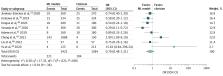
          Of 39 studies that met all criteria and were included in this analysis, 18 (46.2%) used AI models to diagnose hip fractures on plain radiographs and 21 (53.8%) used AI models to predict patient outcomes following hip fracture surgery. A total of 39 598 plain radiographs and 714 939 hip fractures were used for training, validating, and testing ML models specific to diagnosis and postoperative outcome prediction, respectively. Mortality and length of hospital stay were the most predicted outcomes. On pooled data analysis, compared with clinicians, the OR for diagnostic error of ML models was 0.79 (95% CI, 0.48-1.31; P = .36; I 2 = 60%) for hip fracture radiographs. For the ML models, the mean (SD) sensitivity was 89.3% (8.5%), specificity was 87.5% (9.9%), and F1 score was 0.90 (0.06). The mean area under the curve for mortality prediction was 0.84 with ML models compared with 0.79 for alternative controls ( P = .09).

          Conclusions and Relevance

          The findings of this systematic review and meta-analysis suggest that the potential applications of AI to aid with diagnosis from hip radiographs are promising. The performance of AI in diagnosing hip fractures was comparable with that of expert radiologists and surgeons. However, current implementations of AI for outcome prediction do not seem to provide substantial benefit over traditional multivariable predictive statistics.

          Related collections

          Most cited references71

          • Record: found
          • Abstract: found
          • Article: found
          Is Open Access

          The PRISMA 2020 statement: an updated guideline for reporting systematic reviews

          The Preferred Reporting Items for Systematic reviews and Meta-Analyses (PRISMA) statement, published in 2009, was designed to help systematic reviewers transparently report why the review was done, what the authors did, and what they found. Over the past decade, advances in systematic review methodology and terminology have necessitated an update to the guideline. The PRISMA 2020 statement replaces the 2009 statement and includes new reporting guidance that reflects advances in methods to identify, select, appraise, and synthesise studies. The structure and presentation of the items have been modified to facilitate implementation. In this article, we present the PRISMA 2020 27-item checklist, an expanded checklist that details reporting recommendations for each item, the PRISMA 2020 abstract checklist, and the revised flow diagrams for original and updated reviews.
            Bookmark
            • Record: found
            • Abstract: found
            • Article: not found

            Deep learning.

            Deep learning allows computational models that are composed of multiple processing layers to learn representations of data with multiple levels of abstraction. These methods have dramatically improved the state-of-the-art in speech recognition, visual object recognition, object detection and many other domains such as drug discovery and genomics. Deep learning discovers intricate structure in large data sets by using the backpropagation algorithm to indicate how a machine should change its internal parameters that are used to compute the representation in each layer from the representation in the previous layer. Deep convolutional nets have brought about breakthroughs in processing images, video, speech and audio, whereas recurrent nets have shone light on sequential data such as text and speech.
              Bookmark
              • Record: found
              • Abstract: found
              • Article: not found

              Artificial intelligence in radiology

              Artificial intelligence (AI) algorithms, particularly deep learning, have demonstrated remarkable progress in image-recognition tasks. Methods ranging from convolutional neural networks to variational autoencoders have found myriad applications in the medical image analysis field, propelling it forward at a rapid pace. Historically, in radiology practice, trained physicians visually assessed medical images for the detection, characterization and monitoring of diseases. AI methods excel at automatically recognizing complex patterns in imaging data and providing quantitative, rather than qualitative, assessments of radiographic characteristics. In this O pinion article, we establish a general understanding of AI methods, particularly those pertaining to image-based tasks. We explore how these methods could impact multiple facets of radiology, with a general focus on applications in oncology, and demonstrate ways in which these methods are advancing the field. Finally, we discuss the challenges facing clinical implementation and provide our perspective on how the domain could be advanced.
                Bookmark

                Author and article information

                Journal
                JAMA Netw Open
                JAMA Netw Open
                JAMA Network Open
                American Medical Association
                2574-3805
                17 March 2023
                March 2023
                17 March 2023
                : 6
                : 3
                : e233391
                Affiliations
                [1 ]Division of Orthopaedic Surgery, Department of Surgery, University of Toronto, Toronto, Ontario, Canada
                [2 ]Institute of Biomedical Engineering, University of Toronto, Toronto, Ontario, Canada
                [3 ]Orthopaedics Biomechanics Laboratory, Sunnybrook Research Institute, Toronto, Ontario, Canada
                [4 ]Temerty Faculty of Medicine, University of Toronto, Toronto, Ontario, Canada
                [5 ]Division of Orthopaedic Surgery, Sunnybrook Health Sciences Centre, Toronto, Ontario, Canada
                Author notes
                Article Information
                Accepted for Publication: January 31, 2023.
                Published: March 17, 2023. doi:10.1001/jamanetworkopen.2023.3391
                Open Access: This is an open access article distributed under the terms of the CC-BY License. © 2023 Lex JR et al. JAMA Network Open.
                Corresponding Author: Johnathan R. Lex, MB, ChB, Division of Orthopaedic Surgery, Department of Surgery, University of Toronto, 500 University, Room 602, Toronto, ON M5G 1V7, Canada ( johnathanlex@ 123456gmail.com ).
                Author Contributions: Drs Lex and Ravi had full access to all of the data in the study and take responsibility for the integrity of the data and the accuracy of the data analysis.
                Concept and design: Lex, Koucheki, Whyne, Ravi.
                Acquisition, analysis, or interpretation of data: Lex, Di Michele, Koucheki, Pincus.
                Drafting of the manuscript: Lex, Di Michele, Koucheki, Ravi.
                Critical revision of the manuscript for important intellectual content: Di Michele, Koucheki, Pincus, Whyne.
                Statistical analysis: Koucheki.
                Administrative, technical, or material support: Koucheki.
                Supervision: Lex, Di Michele, Pincus, Whyne, Ravi.
                Conflict of Interest Disclosures: Dr Lex reported receiving grants from Arthrex Inc outside the submitted work and serving on the Resident Advisory Board for PrecisionOS Technologies. No other disclosures were reported.
                Funding/Support: Scholarship support for this project (specifically for Dr Lex) was provided by the William and Suzanne Holland Chair in Musculoskeletal Research and the Queen Elizabeth II/Patty Rigby & John Wedge Graduate Scholarships in Science and Technology.
                Role of the Funder/Sponsor: The funding sources had no role in the design and conduct of the study; collection, management, analysis, and interpretation of the data; preparation, review, or approval of the manuscript; and decision to submit the manuscript for publication.
                Data Sharing Statement: See Supplement 2.
                Article
                zoi230135
                10.1001/jamanetworkopen.2023.3391
                10024206
                36930153
                664a10c0-3f56-4de8-acbc-c1b3717e8b06
                Copyright 2023 Lex JR et al. JAMA Network Open.

                This is an open access article distributed under the terms of the CC-BY License.

                History
                : 22 October 2022
                : 31 January 2023
                Categories
                Research
                Original Investigation
                Online Only
                Orthopedics

                Comments

                Comment on this article