3
views
0
recommends
+1 Recommend
0 collections
    0
    shares
      • Record: found
      • Abstract: found
      • Article: found
      Is Open Access

      New microvascular ultrasound techniques: abdominal applications

      review-article

      Read this article at

      Bookmark
          There is no author summary for this article yet. Authors can add summaries to their articles on ScienceOpen to make them more accessible to a non-specialist audience.

          Abstract

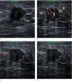
          Microvascular ultrasound (MVUS) is a new ultrasound technique that allows the detection of slow-velocity flow, providing the visualization of the blood flow in small vessels without the need of intravenous contrast agent administration. This technology has been integrated in the most recent ultrasound equipment and applied for the assessment of vascularization. Compared to conventional color Doppler and power Doppler imaging, MVUS provides higher capability to detect intralesional flow. A growing number of studies explored the potential applications in hepatobiliary, genitourinary, and vascular pathologies. Different flow patterns can be observed in hepatic and renal focal lesions providing information on tumor vascularity and improving the differential diagnosis. This article aims to provide a detailed review on the current evidences and applications of MVUS in abdominal imaging.

          Related collections

          Most cited references69

          • Record: found
          • Abstract: found
          • Article: not found

          Liver fibrosis: Review of current imaging and MRI quantification techniques.

          Liver fibrosis is characterized by the accumulation of extracellular matrix proteins such as collagen in the liver interstitial space. All causes of chronic liver disease may lead to fibrosis and cirrhosis. The severity of liver fibrosis influences the decision to treat or the need to monitor hepatic or extrahepatic complications. The traditional reference standard for diagnosis of liver fibrosis is liver biopsy. However, this technique is invasive, associated with a risk of sampling error, and has low patient acceptance. Imaging techniques offer the potential for noninvasive diagnosis, staging, and monitoring of liver fibrosis. Recently, several of these have been implemented on ultrasound (US), computed tomography, or magnetic resonance imaging (MRI). Techniques that assess changes in liver morphology, texture, or perfusion that accompany liver fibrosis have been implemented on all three imaging modalities. Elastography, which measures changes in mechanical properties associated with liver fibrosis-such as strain, stiffness, or viscoelasticity-is available on US and MRI. Some techniques assessing liver shear stiffness have been adopted clinically, whereas others assessing strain or viscoelasticity remain investigational. Further, some techniques are only available on MRI-such as spin-lattice relaxation time in the rotating frame (T1 ρ), diffusion of water molecules, and hepatocellular function based on the uptake of a liver-specific contrast agent-remain investigational in the setting of liver fibrosis staging. In this review, we summarize the key concepts, advantages and limitations, and diagnostic performance of each technique. The use of multiparametric MRI techniques offers the potential for comprehensive assessment of chronic liver disease severity.
            Bookmark
            • Record: found
            • Abstract: found
            • Article: found
            Is Open Access

            Up-to-date Doppler techniques for breast tumor vascularity: superb microvascular imaging and contrast-enhanced ultrasound

            Ah Park, Bo Seo (2017)
            Ultrasonographic Doppler techniques have improved greatly over the years, allowing more sophisticated evaluation of breast tumor vascularity. Superb microvascular imaging (SMI) and contrast-enhanced ultrasound (CEUS) with second-generation contrast agents are two representative up-to-date techniques. SMI is a sensitive Doppler technique that adopts an intelligent filter system to separate low-flow signals from artifacts. With the development of second-generation contrast agents, CEUS has also emerged as a useful Doppler technique for evaluating tumor microcirculation. Both techniques can improve the diagnostic performance of gray-scale ultrasonography by providing vascular information useful not only for the morphologic assessment of microvessels, but also for the quantitative analysis of perfusion. In this review, we explain the imaging principles and previous research underlying these two vascular techniques, and describe our clinical experiences.
              Bookmark
              • Record: found
              • Abstract: found
              • Article: not found

              Clinical Applications of Superb Microvascular Imaging in the Liver, Breast, Thyroid, Skeletal Muscle, and Carotid Plaques.

              This article reviews the clinical applications of Superb Microvascular Imaging (SMI; Canon Medical Systems, Otawara, Japan) in the liver, breast, thyroid, skeletal muscle, and carotid plaques. Diseases that are closely associated with angiogenesis can be diagnosed by SMI in a relatively early phase, and using SMI can prevent adverse reactions associated with the contrast agents used in contrast-enhanced ultrasound. Super Microvascular Imaging also shows particular value in grading disease activities and monitoring therapeutic responses. Although SMI has some limitations, such as a lack of clinical standards, it can add information to conventional ultrasound examinations and may become a noninvasive alternative to invasive diagnostic procedures for many clinical conditions.
                Bookmark

                Author and article information

                Contributors
                roberto.cannella@unipa.it
                Journal
                Radiol Med
                Radiol Med
                La Radiologia Medica
                Springer Milan (Milan )
                0033-8362
                1826-6983
                26 July 2023
                26 July 2023
                2023
                : 128
                : 9
                : 1023-1034
                Affiliations
                GRID grid.10776.37, ISNI 0000 0004 1762 5517, Department of Biomedicine, Neuroscience and Advanced Diagnostics (BiND), , University of Palermo, ; Via del Vespro 129, 90127 Palermo, Italy
                Author information
                http://orcid.org/0000-0002-3808-0785
                http://orcid.org/0000-0002-8808-379X
                Article
                1679
                10.1007/s11547-023-01679-6
                10473992
                37495910
                65e72001-5b08-450e-a821-40126d863e72
                © The Author(s) 2023

                Open Access This article is licensed under a Creative Commons Attribution 4.0 International License, which permits use, sharing, adaptation, distribution and reproduction in any medium or format, as long as you give appropriate credit to the original author(s) and the source, provide a link to the Creative Commons licence, and indicate if changes were made. The images or other third party material in this article are included in the article's Creative Commons licence, unless indicated otherwise in a credit line to the material. If material is not included in the article's Creative Commons licence and your intended use is not permitted by statutory regulation or exceeds the permitted use, you will need to obtain permission directly from the copyright holder. To view a copy of this licence, visit http://creativecommons.org/licenses/by/4.0/.

                History
                : 31 March 2023
                : 3 July 2023
                Funding
                Funded by: Università degli Studi di Palermo
                Categories
                Abdominal Radiology
                Custom metadata
                © Italian Society of Medical Radiology 2023

                superb microvascular imaging,microvascular flow imaging,micro-flow imaging,contrast-enhanced ultrasound

                Comments

                Comment on this article