9
views
0
recommends
+1 Recommend
0 collections
    0
    shares
      • Record: found
      • Abstract: found
      • Article: found
      Is Open Access

      Uncemented three-dimensional-printed prosthetic replacement for giant cell tumor of distal radius: a new design of prosthesis and surgical techniques

      research-article

      Read this article at

      Bookmark
          There is no author summary for this article yet. Authors can add summaries to their articles on ScienceOpen to make them more accessible to a non-specialist audience.

          Abstract

          Introduction

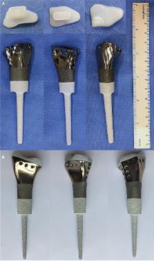
          Currently, it is challenging to treat giant cell tumor (GCT) of distal radius. For Campanacci grade III or recurrent GCTs, en bloc resection has been accepted as a better treatment option. Although numerous methods are available for reconstruction, all of them have some limitations in joint function and complications. In this study, our aims were to treat the GCT of distal radius with uncemented three-dimensional (3D)-printed prosthesis and to present and evaluate the surgical techniques and short-term outcomes.

          Methods

          Between September 2015 and March 2017, 11 patients with distal radius GCTs were treated with personalized uncemented 3D-printed prosthesis. The preoperative/postoperative pain, range of motion, and grip strengths of all patients were evaluated. Oncological results, complications, and degenerative changes in the wrist joint were evaluated. Functional outcomes were assessed according to the disabilities of the arm, shoulder, and hand (DASH) questionnaire and Mayo wrist scoring systems.

          Results

          The average follow-up was 14.45 months (range, 8–18 months). There was a significant decrease in the mean postoperative visual analog scale score (2.33) compared with the preoperative score (5.22; p<0.001). The mean DASH score and Mayo wrist score of the wrist joint function were 18.7 and 72, respectively. There was no local recurrence or lung metastasis. No complication associated with prosthesis was observed, including aseptic loosening, subluxation, and breakage. Joint space narrowing, or disuse osteoporosis, was also not found in all cases.

          Conclusion

          En bloc resection and reconstruction with a personalized uncemented 3D-printed prosthesis can be alternative options to treat Campanacci grade III or recurrent GCTs of distal radius and can result in short-term oncologic salvage, good postoperative function, and low complication rate. However, a long-term follow-up is required to determine the outcome.

          Related collections

          Most cited references24

          • Record: found
          • Abstract: found
          • Article: not found

          Giant-cell tumor of bone.

          Of 327 patients who had a giant-cell tumor of bone and were seen at the Istituto Rizzoli, 293 were treated at the Institute, and 280 of these were followed for two to forty-four years. The distribution according to sex and age of the patient and site of the tumor was similar to the distributions in major reports of large series. The tumor usually involved the metaphysis and the epiphysis, but was occasionally limited to the metaphysis, and in only 2 per cent of the patients was it adjacent to an open growth plate. The tumor on occasion invaded the articular space, also involving the ligaments and the synovial membrane. Extension to an adjacent bone through the joint occurred in 5 per cent of the tumors. Our radiographic grading, which is roughly comparable with the staging system of Enneking et al., was Grade I in 4 per cent, II in 74 per cent, and III in 22 per cent of 266 patients before treatment. A pathological fracture was apparent on the first radiograph in 9 per cent of the patients. In the 280 patients with adequate follow-up, 331 surgical procedures were performed. The rate of local recurrence was 27 per cent in the 151 intralesional procedures, 8 per cent in the 122 marginal excisions, and zero in the fifty-eight wide or radical procedures. These results did not correlate with the radiographic grade of the lesion. Of the fifty-one local recurrences that were seen after treatment at our institution, 90 per cent appeared in the first three years after surgery.(ABSTRACT TRUNCATED AT 250 WORDS)
            Bookmark
            • Record: found
            • Abstract: not found
            • Article: not found

            Giant-cell tumor of bone. An analysis of two hundred and eighteen cases.

              Bookmark
              • Record: found
              • Abstract: found
              • Article: not found

              Giant cell tumor of bone.

              Giant cell tumor (GCT) of bone is one type of giant cell-rich lesion of bone. This benign mesenchymal tumor has characteristic multinuclear giant cells. Mononuclear stromal cells are the physiologically active and diagnostic cell type. Most GCTs are located in the epiphyseal regions of long bones. The axial skeleton-primarily the sacrum-is a secondary site of involvement. Most patients present with pain, swelling, joint effusion, and disability in the third and fourth decades of life. Imaging studies are important for tumor staging and radiographic grading. Typically, these clinically active but slow-growing tumors are confined to bone, with relatively well-defined radiographic borders. Monostotic disease is most common. Metastatic spread to the lungs is rare. Extended intralesional curettage with or without adjuvant therapy is the primary treatment choice. Local recurrence is seen in ≤ 20% of cases, and a second local intralesional procedure is typically sufficient in cases that are detected early. Medical therapies include diphosphonates and denosumab. Denosumab has been approved for use in osteoporosis as well as breast and prostate cancer metastatic to bone. Medical therapy and radiotherapy can alter the management of GCT of bone, especially in multifocal disease, local recurrences, and bulky central/axial disease.
                Bookmark

                Author and article information

                Journal
                Cancer Manag Res
                Cancer Manag Res
                Cancer Management and Research
                Cancer Management and Research
                Dove Medical Press
                1179-1322
                2018
                08 February 2018
                : 10
                : 265-277
                Affiliations
                Department of Orthopedics, West China Hospital, Sichuan University, Chengdu, Sichuan, People’s Republic of China
                Author notes
                Correspondence: Chongqi Tu, Department of Orthopedics, West China Hospital, Sichuan University, No. 37 Guoxue Street, Chengdu 610041, Sichuan, People’s Republic of China, Tel +86 182 8006 0169, Fax +86 028 8542 2454, Email Tuchongqi@ 123456yeah.net
                [*]

                These authors contributed equally to this work

                Article
                cmar-10-265
                10.2147/CMAR.S146434
                5808706
                29445303
                654d0324-829c-4cc2-8199-1356a9f82ba5
                © 2018 Lu et al. This work is published and licensed by Dove Medical Press Limited

                The full terms of this license are available at https://www.dovepress.com/terms.php and incorporate the Creative Commons Attribution – Non Commercial (unported, v3.0) License ( http://creativecommons.org/licenses/by-nc/3.0/). By accessing the work you hereby accept the Terms. Non-commercial uses of the work are permitted without any further permission from Dove Medical Press Limited, provided the work is properly attributed.

                History
                Categories
                Original Research

                Oncology & Radiotherapy
                rapid prototyping,computer-aided design,cementless,uncemented,polyethylene liner

                Comments

                Comment on this article