3
views
0
recommends
+1 Recommend
0 collections
    0
    shares
      • Record: found
      • Abstract: found
      • Article: found
      Is Open Access

      Optimized Surface Characteristics and Enhanced in Vivo Osseointegration of Alkali-Treated Titanium with Nanonetwork Structures

      research-article

      Read this article at

      Bookmark
          There is no author summary for this article yet. Authors can add summaries to their articles on ScienceOpen to make them more accessible to a non-specialist audience.

          Abstract

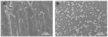
          Alkali-treated titanium (Ti) with a porous, homogeneous, and uniform nanonetwork structure (TNS) that enables establishment of a more rapid and firmer osteointegration than titanium has recently been reported. However, the mechanisms underlying the enhanced osteogenic activity on TNS remains to be elucidated. This study aimed to evaluate the surface physicochemical properties of Ti and TNS, and investigate osteoinduction and osteointegration in vivo. Surface characteristics were evaluated using scanning electron microscopy (SEM), scanning probe microscopy (SPM), and X-ray photoelectron spectrometry (XPS), and the surface electrostatic force of TNS was determined using solid zeta potential. This study also evaluated the adsorption of bovine serum albumin (BSA) and human plasma fibronectin (HFN) on Ti and TNS surfaces using quartz crystal microbalance (QCM) sensors, and apatite formation on Ti and TNS surfaces was examined using a simulated body fluid (SBF) test. Compared with Ti, the newly developed TNS enhanced BSA and HFN absorbance capacity and promoted apatite formation. Furthermore, TNS held less negative charge than Ti. Notably, sequential fluorescence labeling and microcomputed tomography assessment indicated that TNS screws implanted into rat femurs exhibited remarkably enhanced osteointegration compared with Ti screws. These results indicate that alkali-treated titanium implant with a nanonetwork structure has considerable potential for future clinical applications in dentistry and orthopedics.

          Related collections

          Most cited references42

          • Record: found
          • Abstract: not found
          • Article: not found

          Ti based biomaterials, the ultimate choice for orthopaedic implants – A review

            Bookmark
            • Record: found
            • Abstract: found
            • Article: not found

            Surface treatments of titanium dental implants for rapid osseointegration.

            The osseointegration rate of titanium dental implants is related to their composition and surface roughness. Rough-surfaced implants favor both bone anchoring and biomechanical stability. Osteoconductive calcium phosphate coatings promote bone healing and apposition, leading to the rapid biological fixation of implants. The different methods used for increasing surface roughness or applying osteoconductive coatings to titanium dental implants are reviewed. Surface treatments, such as titanium plasma-spraying, grit-blasting, acid-etching, anodization or calcium phosphate coatings, and their corresponding surface morphologies and properties are described. Most of these surfaces are commercially available and have proven clinical efficacy (>95% over 5 years). The precise role of surface chemistry and topography on the early events in dental implant osseointegration remain poorly understood. In addition, comparative clinical studies with different implant surfaces are rarely performed. The future of dental implantology should aim to develop surfaces with controlled and standardized topography or chemistry. This approach will be the only way to understand the interactions between proteins, cells and tissues, and implant surfaces. The local release of bone stimulating or resorptive drugs in the peri-implant region may also respond to difficult clinical situations with poor bone quality and quantity. These therapeutic strategies should ultimately enhance the osseointegration process of dental implants for their immediate loading and long-term success.
              Bookmark
              • Record: found
              • Abstract: found
              • Article: not found

              Solutions able to reproduce in vivo surface-structure changes in bioactive glass-ceramic A-W.

              High-strength bioactive glass-ceramic A-W was soaked in various acellular aqueous solutions different in ion concentrations and pH. After soaking for 7 and 30 days, surface structural changes of the glass-ceramic were investigated by means of Fourier transform infrared reflection spectroscopy, thin-film x-ray diffraction, and scanning electronmicroscopic observations, in comparison with in vivo surface structural changes. So-called Tris buffer solution, pure water buffered with trishydroxymethyl-aminomethane, which had been used by various workers as a "simulated body fluid," did not reproduce the in vivo surface structural changes, i.e., apatite formation on the surface. A solution, ion concentrations and pH of which are almost equal to those of the human blood plasma--i.e., Na+ 142.0, K+ 5.0, Mg2+ 1.5, Ca2+ 2.5, Cl- 148.8, HCO3- 4.2 and PO4(2-) 1.0 mM and buffered at pH 7.25 with the trishydroxymethyl-aminomethane--most precisely reproduced in vivo surface structure change. This shows that careful selection of simulated body fluid is required for in vitro experiments. The results also support the concept that the apatite phase on the surface of glass-ceramic A-W is formed by a chemical reaction of the glass-ceramic with the Ca2+, HPO4(2-), and OH- ions in the body fluid.
                Bookmark

                Author and article information

                Journal
                Int J Mol Sci
                Int J Mol Sci
                ijms
                International Journal of Molecular Sciences
                MDPI
                1422-0067
                05 March 2019
                March 2019
                : 20
                : 5
                : 1127
                Affiliations
                [1 ]Department of Removable Prosthodontics and Occlusion, Osaka Dental University, 8-1, Kuzuhahanazono-cho, Hirakata-shi, Osaka 573-1121, Japan; zeng-yuhao@ 123456hotmail.com (Y.Z.); yangyuanyuan0801@ 123456outlook.com (Y.Y.); y.d.r.nld@ 123456hotmail.com (D.Y.); joecheung_asuka@ 123456hotmail.com (H.Z.); tashiro@ 123456cc.osaka-dent.ac.jp (Y.T.); clair.de.lune.peridota@ 123456gmail.com (S.I.); joji@ 123456cc.osaka-dent.ac.jp (J.O.); komasa-s@ 123456cc.osaka-dent.ac.jp (S.K.)
                [2 ]Faculty of Health Sciences, Osaka Dental University, 1-4-4, Makino-honmachi, Hirakata-shi, Osaka 573-1144, Japan; kusumoto@ 123456cc.osaka-dent.ac.jp (T.K.); nisizaki@ 123456cc.osaka-dent.ac.jp (H.N.)
                [3 ]The Institute of Scientific and Industrial Research, Osaka University, Suita, Osaka 565-0871, Japan; sekino@ 123456sanken.osaka-u.ac.jp
                Author notes
                [* ]Correspondence: chen-luyuan900115@ 123456foxmail.com ; Tel.: +81-080-3824-7328
                [†]

                These authors contributed equally to this work.

                Author information
                https://orcid.org/0000-0001-6778-6915
                https://orcid.org/0000-0002-6605-9166
                https://orcid.org/0000-0001-5400-8479
                Article
                ijms-20-01127
                10.3390/ijms20051127
                6429490
                30841636
                64cc64e9-2c3c-4f2d-b768-c095ea8de275
                © 2019 by the authors.

                Licensee MDPI, Basel, Switzerland. This article is an open access article distributed under the terms and conditions of the Creative Commons Attribution (CC BY) license ( http://creativecommons.org/licenses/by/4.0/).

                History
                : 10 January 2019
                : 28 February 2019
                Categories
                Article

                Molecular biology
                implant,alkali treatment,in vivo study,nanonetwork,osseointegration
                Molecular biology
                implant, alkali treatment, in vivo study, nanonetwork, osseointegration

                Comments

                Comment on this article