3
views
0
recommends
+1 Recommend
0 collections
    0
    shares
      • Record: found
      • Abstract: found
      • Article: found
      Is Open Access

      Fetal myocardial deformation measured with two‐dimensional speckle‐tracking echocardiography: longitudinal prospective cohort study of 124 healthy fetuses

      research-article

      Read this article at

      Bookmark
          There is no author summary for this article yet. Authors can add summaries to their articles on ScienceOpen to make them more accessible to a non-specialist audience.

          ABSTRACT

          Objectives

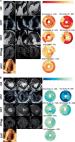
          Two‐dimensional speckle‐tracking echocardiography (2D‐STE) is a promising technique which allows assessment of fetal cardiac function, and can be used in the evaluation of cardiac and non‐cardiac diseases in pregnancy. However, reliable fetal reference values for deformation parameters measured using 2D‐STE are needed before it can be introduced into clinical practice. This study aimed to obtain reference values for fetal global longitudinal strain (GLS) and GLS rate (GLSR) measured using 2D‐STE and compare right and left ventricular values.

          Methods

          This was a prospective longitudinal cohort study of uncomplicated pregnancies that underwent echocardiography every 4 weeks from inclusion at 18–21 weeks until delivery to obtain four‐chamber loops of the fetal heart. Left and right ventricular GLS and GLSR were measured using 2D‐STE at each examination. Using Bayesian mixed‐effects models, reference values with lower and upper 5% prediction limits were calculated according to gestational age. Right and left ventricular GLS values according to gestational age were compared using the Wilcoxon signed‐rank test.

          Results

          A total of 592 left ventricular and 566 right ventricular GLS and GLSR measurements were obtained from 124 women with uncomplicated pregnancy and non‐anomalous, appropriately grown fetuses. Reference values were obtained for both fetal ventricles according to gestational week. GLS and GLSR values of both ventricles increased (i.e. became less negative) significantly during pregnancy. Right ventricular GLS values were significantly higher (i.e. less negative) than the respective left ventricular values at every gestational week.

          Conclusions

          Reference values were obtained for fetal GLS and GLSR measured using 2D‐STE. GLS and GLSR values increased significantly for both ventricles from the second trimester until delivery. GLS values were significantly higher for the right ventricle compared with the left ventricle. Future studies are needed to assess whether the obtained reference values are helpful in clinical practice in the assessment of pregnancy complications, such as fetal growth restriction or cardiac anomaly. © 2022 The Authors. Ultrasound in Obstetrics & Gynecology published by John Wiley & Sons Ltd on behalf of International Society of Ultrasound in Obstetrics and Gynecology.

          Related collections

          Most cited references69

          • Record: found
          • Abstract: found
          • Article: not found

          Definitions for a common standard for 2D speckle tracking echocardiography: consensus document of the EACVI/ASE/Industry Task Force to standardize deformation imaging.

          Recognizing the critical need for standardization in strain imaging, in 2010, the European Association of Echocardiography (now the European Association of Cardiovascular Imaging, EACVI) and the American Society of Echocardiography (ASE) invited technical representatives from all interested vendors to participate in a concerted effort to reduce intervendor variability of strain measurement. As an initial product of the work of the EACVI/ASE/Industry initiative to standardize deformation imaging, we prepared this technical document which is intended to provide definitions, names, abbreviations, formulas, and procedures for calculation of physical quantities derived from speckle tracking echocardiography and thus create a common standard.
            Bookmark
            • Record: found
            • Abstract: found
            • Book: not found

            Practical Statistics for Medical Research

            Most medical researchers, whether clinical or non-clinical, receive some background in statistics as undergraduates. However, it is most often brief, a long time ago, and largely forgotten by the time it is needed. Furthermore, many introductory texts fall short of adequately explaining the underlying concepts of statistics, and often are divorced from the reality of conducting and assessing medical research.<br><br>Practical Statistics for Medical Research is a problem-based text for medical researchers, medical students, and others in the medical arena who need to use statistics but have no specialized mathematics background. <br><br>The author draws on twenty years of experience as a consulting medical statistician to provide clear explanations to key statistical concepts, with a firm emphasis on practical aspects of designing and analyzing medical research. The text gives special attention to the presentation and interpretation of results and the many real problems that arise in medical research.
              Bookmark
              • Record: found
              • Abstract: found
              • Article: found
              Is Open Access

              Myocardial strain imaging: review of general principles, validation, and sources of discrepancies

              Abstract Myocardial tissue tracking imaging techniques have been developed for a more accurate evaluation of myocardial deformation (i.e. strain), with the potential to overcome the limitations of ejection fraction (EF) and to contribute, incremental to EF, to the diagnosis and prognosis in cardiac diseases. While most of the deformation imaging techniques are based on the similar principles of detecting and tracking specific patterns within an image, there are intra- and inter-imaging modality inconsistencies limiting the wide clinical applicability of strain. In this review, we aimed to describe the particularities of the echocardiographic and cardiac magnetic resonance deformation techniques, in order to understand the discrepancies in strain measurement, focusing on the potential sources of variation: related to the software used to analyse the data, to the different physics of image acquisition and the different principles of 2D vs. 3D approaches. As strain measurements are not interchangeable, it is highly desirable to work with validated strain assessment tools, in order to derive information from evidence-based data. There is, however, a lack of solid validation of the current tissue tracking techniques, as only a few of the commercial deformation imaging softwares have been properly investigated. We have, therefore, addressed in this review the neglected issue of suboptimal validation of tissue tracking techniques, in order to advocate for this matter.
                Bookmark

                Author and article information

                Contributors
                chantelle.de.vet@mmc.nl
                Journal
                Ultrasound Obstet Gynecol
                Ultrasound Obstet Gynecol
                10.1002/(ISSN)1469-0705
                UOG
                Ultrasound in Obstetrics & Gynecology
                John Wiley & Sons, Ltd. (Chichester, UK )
                0960-7692
                1469-0705
                06 April 2022
                May 2022
                : 59
                : 5 ( doiID: 10.1002/uog.v59.5 )
                : 651-659
                Affiliations
                [ 1 ] Eindhoven MedTech Innovation Centre (e/MTIC) Eindhoven The Netherlands
                [ 2 ] Department of Electrical Engineering Eindhoven University of Technology Eindhoven The Netherlands
                [ 3 ] Department of Gynaecology and Obstetrics Ghent University Ghent Belgium
                [ 4 ] Department of Gynaecology and Obstetrics Máxima Medical Centre Veldhoven The Netherlands
                [ 5 ] Department of Paediatric Cardiology, Emma Children's Hospital, Academic Medical Center Amsterdam University Medical Centers Amsterdam The Netherlands
                [ 6 ] Department of Mathematics & Computer Science Eindhoven University of Technology Eindhoven The Netherlands
                Author notes
                [*] [* ] Correspondence to: Dr C. M. de Vet, Máxima Medical Centre, De Run 4600, 5504 DB Veldhoven, The Netherlands (e‐mail: Chantelle.de.Vet@ 123456mmc.nl )

                N.H.M.v.O. and C.M.d.V. contributed equally to this study.

                Author information
                https://orcid.org/0000-0002-9605-5334
                https://orcid.org/0000-0001-8551-9626
                https://orcid.org/0000-0001-5276-1475
                https://orcid.org/0000-0002-2126-3411
                https://orcid.org/0000-0001-9157-7224
                https://orcid.org/0000-0002-0157-3332
                https://orcid.org/0000-0001-5350-3267
                Article
                UOG24781
                10.1002/uog.24781
                9321172
                34558747
                64ba405e-a8cf-4895-bcef-5471078de5d5
                © 2022 The Authors. Ultrasound in Obstetrics & Gynecology published by John Wiley & Sons Ltd on behalf of International Society of Ultrasound in Obstetrics and Gynecology.

                This is an open access article under the terms of the http://creativecommons.org/licenses/by-nc-nd/4.0/ License, which permits use and distribution in any medium, provided the original work is properly cited, the use is non‐commercial and no modifications or adaptations are made.

                History
                : 14 September 2021
                : 05 March 2021
                : 16 September 2021
                Page count
                Figures: 4, Tables: 4, Pages: 9, Words: 5670
                Categories
                Original Paper
                Original Papers
                Custom metadata
                2.0
                May 2022
                Converter:WILEY_ML3GV2_TO_JATSPMC version:6.1.7 mode:remove_FC converted:26.07.2022

                Obstetrics & Gynecology
                deformation values,fetal strain,pregnancy,strain rate,two‐dimensional speckle tracking

                Comments

                Comment on this article