69
views
0
recommends
+1 Recommend
0 collections
    0
    shares
      • Record: found
      • Abstract: found
      • Article: not found

      Immunosuppressive plasma cells impede T cell-dependent immunogenic chemotherapy

      research-article

      Read this article at

      ScienceOpenPublisherPMC
      Bookmark
          There is no author summary for this article yet. Authors can add summaries to their articles on ScienceOpen to make them more accessible to a non-specialist audience.

          Abstract

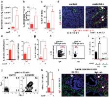
          Cancer-associated genetic alterations induce expression of tumor antigens which can activate CD8 + cytotoxic T cells (CTL), but the microenvironment of established tumors promotes immune tolerance through poorly understood mechanisms 1, 2 . Recently developed therapeutics that overcome tolerogenic mechanisms activate tumor-directed CTL and are effective in some human cancers 1 . Immune mechanisms also affect treatment outcome and certain chemotherapeutic drugs stimulate cancer-specific immune responses by inducing immunogenic cell death (ICD) and other effector mechanisms 3, 4 . Our previous studies revealed that B lymphocytes recruited by CXCL13 into prostate cancer (PC) promote castrate-resistant PC (CRPC) by producing lymphotoxin (LT) which activates an IKKα-Bmi1 module in PC stem cells 5, 6 . Since CRPC is refractory to most therapies, we examined B cell involvement in acquisition of chemotherapy resistance. We focused this study on oxaliplatin, an immunogenic chemotherapeutic 3, 4 that is effective in aggressive PC 7 . We found that B cells modulate the response to low dose oxaliplatin, which by inducing ICD promotes tumor-directed CTL activation. Three different mouse PC models were refractory to oxaliplatin unless genetically or pharmacologically depleted of B cells. The critical immunosuppressive B cells are plasmocytes that express IgA, IL-10 and PD-L1, whose appearance depends on TGFβ-receptor (TGFβR) signaling. Elimination of these cells, which also infiltrate human therapy-resistant PC, allows CTL-dependent eradication of oxaliplatin-treated tumors.

          Related collections

          Most cited references38

          • Record: found
          • Abstract: found
          • Article: not found

          The blockade of immune checkpoints in cancer immunotherapy.

          Among the most promising approaches to activating therapeutic antitumour immunity is the blockade of immune checkpoints. Immune checkpoints refer to a plethora of inhibitory pathways hardwired into the immune system that are crucial for maintaining self-tolerance and modulating the duration and amplitude of physiological immune responses in peripheral tissues in order to minimize collateral tissue damage. It is now clear that tumours co-opt certain immune-checkpoint pathways as a major mechanism of immune resistance, particularly against T cells that are specific for tumour antigens. Because many of the immune checkpoints are initiated by ligand-receptor interactions, they can be readily blocked by antibodies or modulated by recombinant forms of ligands or receptors. Cytotoxic T-lymphocyte-associated antigen 4 (CTLA4) antibodies were the first of this class of immunotherapeutics to achieve US Food and Drug Administration (FDA) approval. Preliminary clinical findings with blockers of additional immune-checkpoint proteins, such as programmed cell death protein 1 (PD1), indicate broad and diverse opportunities to enhance antitumour immunity with the potential to produce durable clinical responses.
            Bookmark
            • Record: found
            • Abstract: found
            • Article: not found

            T cell receptor antagonist peptides induce positive selection.

            We have used organ culture of fetal thymic lobes from T cell receptor (TCR) transgenic beta 2M(-/-) mice to study the role of peptides in positive selection. The TCR used was from a CD8+ T cell specific for ovalbumin 257-264 in the context of Kb. Several peptides with the ability to induce positive selection were identified. These peptide-selected thymocytes have the same phenotype as mature CD8+ T cells and can respond to antigen. Those peptides with the ability to induce positive selection were all variants of the antigenic peptide and were identified as TCR antagonist peptides for this receptor. One peptide tested, E1, induced positive selection on the beta 2M(-/-) background but negative selection on the beta 2M(+/-) background. These results show that the process of positive selection is exquisitely peptide specific and sensitive to extremely low ligand density and support the notion that low efficacy ligands mediate positive selection.
              Bookmark
              • Record: found
              • Abstract: found
              • Article: not found

              Gene expression profiling identifies clinically relevant subtypes of prostate cancer.

              Prostate cancer, a leading cause of cancer death, displays a broad range of clinical behavior from relatively indolent to aggressive metastatic disease. To explore potential molecular variation underlying this clinical heterogeneity, we profiled gene expression in 62 primary prostate tumors, as well as 41 normal prostate specimens and nine lymph node metastases, using cDNA microarrays containing approximately 26,000 genes. Unsupervised hierarchical clustering readily distinguished tumors from normal samples, and further identified three subclasses of prostate tumors based on distinct patterns of gene expression. High-grade and advanced stage tumors, as well as tumors associated with recurrence, were disproportionately represented among two of the three subtypes, one of which also included most lymph node metastases. To further characterize the clinical relevance of tumor subtypes, we evaluated as surrogate markers two genes differentially expressed among tumor subgroups by using immunohistochemistry on tissue microarrays representing an independent set of 225 prostate tumors. Positive staining for MUC1, a gene highly expressed in the subgroups with "aggressive" clinicopathological features, was associated with an elevated risk of recurrence (P = 0.003), whereas strong staining for AZGP1, a gene highly expressed in the other subgroup, was associated with a decreased risk of recurrence (P = 0.0008). In multivariate analysis, MUC1 and AZGP1 staining were strong predictors of tumor recurrence independent of tumor grade, stage, and preoperative prostate-specific antigen levels. Our results suggest that prostate tumors can be usefully classified according to their gene expression patterns, and these tumor subtypes may provide a basis for improved prognostication and treatment stratification.
                Bookmark

                Author and article information

                Journal
                0410462
                6011
                Nature
                Nature
                Nature
                0028-0836
                1476-4687
                24 March 2015
                29 April 2015
                7 May 2015
                07 November 2015
                : 521
                : 7550
                : 94-98
                Affiliations
                [1 ]Laboratory of Gene Regulation and Signal Transduction, Department of Pharmacology, University of California San Diego (UCSD), 9500 Gilman Drive, San Diego CA 92093
                [2 ]Department of Pathology, School of Medicine, University of California San Diego (UCSD), 9500 Gilman Drive, San Diego CA 92093
                [3 ]Institute of Immunology, Max Delbrück Center for Molecular Medicine, Charité Campus Buch, 13125 Berlin, Germany
                [4 ]Department of Surgery, Urology Division, UCSD
                [5 ]Department of Urology, Medical University of Vienna, 1090 Vienna, Austria
                [6 ]Department of Pathology, Medical University of Vienna, 1090 Vienna, Austria
                [7 ]Clinical Institute of Pathology, Ludwig Boltzmann Institute for Cancer Research, Medical University of Vienna, Unit of Pathology of Laboratory Animals (UPLA), University of Veterinary Medicine Vienna, 1210 Vienna, Austria
                Article
                NIHMS670944
                10.1038/nature14395
                4501632
                25924065
                643da9c8-67f9-4f88-a77a-f664780e9381
                History
                Categories
                Article

                Uncategorized
                Uncategorized

                Comments

                Comment on this article