32
views
0
recommends
+1 Recommend
0 collections
    0
    shares
      • Record: found
      • Abstract: found
      • Article: found
      Is Open Access

      Electroactive barium titanate coated titanium scaffold improves osteogenesis and osseointegration with low-intensity pulsed ultrasound for large segmental bone defects

      research-article

      Read this article at

      Bookmark
          There is no author summary for this article yet. Authors can add summaries to their articles on ScienceOpen to make them more accessible to a non-specialist audience.

          Abstract

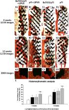
          For large segmental bone defects, porous titanium scaffolds have some advantages, however, they lack electrical activity which hinders their further use. In this study, a barium titanate (BaTiO 3) piezoelectric ceramic was used to modify the surface of a porous Ti6Al4V scaffold (pTi), which was characterized by scanning electron microscopy, energy dispersive spectroscopy, X-ray photoelectron spectroscopy, and roughness and water contact angle analyses. Low intensity pulsed ultrasound (LIPUS) was applied in vitro and in vivo study. The activity of bone marrow mesenchymal stem cells, including adhesion, proliferation, and gene expression, was significantly superior in the BaTiO 3/pTi, pTi + LIPUS, and BaTiO 3/pTi + LIPUS groups than in the pTi group. The activity was also higher in the BaTiO 3/pTi + LIPUS group than in the BaTiO 3/pTi and pTi + LIPUS groups. Additionally, micro-computed tomography, the mineral apposition rate, histomorphology, and the peak pull-out load showed that these scaffold conditions significantly enhanced osteogenesis and osseointegration 6 and 12 weeks after implantation in large segmental bone defects in the radius of rabbits compared with those resulting from the pTi condition. Consequently, the improved osteogenesis and osseointegration make the BaTiO 3/pTi + LIPUS a promising method to promote bone regeneration in large segmental bone defects for clinical application.

          Graphical abstract

          Highlights

          • BaTiO 3 coating was successfully fabricated by a wet chemical method on the surface of porous Ti6Al4V scaffolds.

          • BaTiO 3 coating showed better surface hydrophilicity and roughness than pure porous Ti6Al4V scaffolds.

          • BaTiO 3 coating improved the biological behavior of BMSCs in vitro and promoted the formation of new bones in vivo.

          • LIPUS activated piezoelectric effect of BaTiO 3 coating, enhanced cell viability in vitro and promoted osteogenesis in vivo.

          • BaTiO 3/pTi + LIPUS conditions showed promising potential as a method to repair long bone defects for clinical application.

          Related collections

          Most cited references52

          • Record: found
          • Abstract: not found
          • Article: not found

          Electrospun polymer biomaterials

            Bookmark
            • Record: found
            • Abstract: found
            • Article: not found

            Mechanical evaluation of porous titanium (Ti6Al4V) structures with electron beam melting (EBM).

            Patient specific porous implants for the reconstruction of craniofacial defects have gained importance due to their better performance over their generic counterparts. The recent introduction of electron beam melting (EBM) for the processing of titanium has led to a one step fabrication of porous custom titanium implants with controlled porosity to meet the requirements of the anatomy and functions at the region of implantation. This paper discusses an image based micro-structural analysis and the mechanical characterization of porous Ti6Al4V structures fabricated using the EBM rapid manufacturing process. SEM studies have indicated the complete melting of the powder material with no evidence of poor inter-layer bonding. Micro-CT scan analysis of the samples indicate well formed titanium struts and fully interconnected pores with porosities varying from 49.75%-70.32%. Compression tests of the samples showed effective stiffness values ranging from 0.57(+/-0.05)-2.92(+/-0.17)GPa and compressive strength values of 7.28(+/-0.93)-163.02(+/-11.98)MPa. For nearly the same porosity values of 49.75% and 50.75%, with a variation in only the strut thickness in the sample sets, the compressive stiffness and strength decreased significantly from 2.92 GPa to 0.57 GPa (80.5% reduction) and 163.02 MPa to 7.28 MPa (93.54 % reduction) respectively. The grain density of the fabricated Ti6Al4V structures was found to be 4.423 g/cm(3) equivalent to that of dense Ti6Al4V parts fabricated using conventional methods. In conclusion, from a mechanical strength viewpoint, we have found that the porous structures produced by the electron beam melting process present a promising rapid manufacturing process for the direct fabrication of customized titanium implants for enabling personalized medicine. (c) 2009 Elsevier Ltd. All rights reserved.
              Bookmark
              • Record: found
              • Abstract: found
              • Article: not found

              The critical size defect as an experimental model for craniomandibulofacial nonunions.

              Little consistency has been manifest among investigators in choosing an appropriate experimental model for maxillofacial bone research. In an effort to develop a protocol for the experimental analysis of maxillofacial nonunions, previous studies using calvarial and mandibular defects as models were reviewed. The creation of nonunions in animals within the calvaria and mandible was size dependent. Defects of a size that will not heal during the lifetime of the animal may be termed critical size defects (CSDs). A rationale was postulated for testing bone repair materials (BRMs) using CSDs in a hierarchy of animal models. This rationale suggests that testing should be initiated in the calvaria of the rat and rabbit, followed by testing in the mandibles of dogs and monkeys. While calvarial CSDs have been established in the rat, rabbit, and dog, further research is necessary to determine the CSD in the calvaria of the monkey, as well as the mandibles of dogs and monkeys.
                Bookmark

                Author and article information

                Contributors
                Journal
                Bioact Mater
                Bioact Mater
                Bioactive Materials
                KeAi Publishing
                2452-199X
                15 July 2020
                December 2020
                15 July 2020
                : 5
                : 4
                : 1087-1101
                Affiliations
                [a ]Department of Orthopedics, Xijing Hospital, Fourth Military Medical University, Xi'an, 710032, China
                [b ]Orthopedic Centre-Spine Surgery, The 940 Hospital of Joint Logistics Support Force of Chinese People's Liberation Army, Lanzhou, 730050, China
                [c ]Department of Joint Surgery and Sports Medicine, Hunan Provincial People's Hospital and The First Affiliated Hospital of Hunan Normal University, Changsha, 410016, PR China
                [d ]Shenyang National Laboratory for Materials Science, Institute of Metal Research, Chinese Academy of Sciences, Shenyang, 110016, China
                Author notes
                []Corresponding author. guozheng@ 123456fmmu.edu.cn
                [1]

                These authors contributed equally to this work.

                Article
                S2452-199X(20)30128-6
                10.1016/j.bioactmat.2020.07.001
                7363989
                32695938
                63c1ff82-06a0-4c00-a55a-e86438999779
                © 2020 The Authors. Publishing services by Elsevier B.V. on behalf of KeAi Communications Co., Ltd.

                This is an open access article under the CC BY-NC-ND license (http://creativecommons.org/licenses/by-nc-nd/4.0/).

                History
                : 19 May 2020
                : 3 July 2020
                : 3 July 2020
                Categories
                Article

                large segmental bone defect,barium titanate,piezoelectric ceramic,porous ti6al4v scaffold,osteogenesis

                Comments

                Comment on this article