1
views
0
recommends
+1 Recommend
0 collections
    0
    shares
      • Record: found
      • Abstract: found
      • Article: found
      Is Open Access

      Arrangement and symmetry of the fungal E3BP-containing core of the pyruvate dehydrogenase complex

      research-article

      Read this article at

      Bookmark
          There is no author summary for this article yet. Authors can add summaries to their articles on ScienceOpen to make them more accessible to a non-specialist audience.

          Abstract

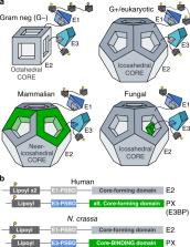
          The pyruvate dehydrogenase complex (PDC) is a multienzyme complex central to aerobic respiration, connecting glycolysis to mitochondrial oxidation of pyruvate. Similar to the E3-binding protein (E3BP) of mammalian PDC, PX selectively recruits E3 to the fungal PDC, but its divergent sequence suggests a distinct structural mechanism. Here, we report reconstructions of PDC from the filamentous fungus Neurospora crassa by cryo-electron microscopy, where we find protein X (PX) interior to the PDC core as opposed to substituting E2 core subunits as in mammals. Steric occlusion limits PX binding, resulting in predominantly tetrahedral symmetry, explaining previous observations in Saccharomyces cerevisiae. The PX-binding site is conserved in (and specific to) fungi, and complements possible C-terminal binding motifs in PX that are absent in mammalian E3BP. Consideration of multiple symmetries thus reveals a differential structural basis for E3BP-like function in fungal PDC.

          Abstract

          The pyruvate dehydrogenase complex (PDC) is a multienzyme complex connecting glycolysis to mitochondrial oxidation of pyruvate. Cryo-EM analysis of PDC from Neurospora crassa reveals localization of fungi-specific protein X (PX) and confirms that it functions like the mammalian E3BP, recruiting the E3 component of PDC.

          Related collections

          Most cited references36

          • Record: found
          • Abstract: found
          • Article: not found

          Regulation of pyruvate metabolism and human disease

          Pyruvate is a keystone molecule critical for numerous aspects of eukaryotic and human metabolism. Pyruvate is the end-product of glycolysis, is derived from additional sources in the cellular cytoplasm, and is ultimately destined for transport into mitochondria as a master fuel input undergirding citric acid cycle carbon flux. In mitochondria, pyruvate drives ATP production by oxidative phosphorylation and multiple biosynthetic pathways intersecting the citric acid cycle. Mitochondrial pyruvate metabolism is regulated by many enzymes, including the recently discovered mitochondria pyruvate carrier, pyruvate dehydrogenase, and pyruvate carboxylase, to modulate overall pyruvate carbon flux. Mutations in any of the genes encoding for proteins regulating pyruvate metabolism may lead to disease. Numerous cases have been described. Aberrant pyruvate metabolism plays an especially prominent role in cancer, heart failure, and neurodegeneration. Because most major diseases involve aberrant metabolism, understanding and exploiting pyruvate carbon flux may yield novel treatments that enhance human health.
            Bookmark
            • Record: found
            • Abstract: found
            • Article: not found

            A key role for mitochondrial gatekeeper pyruvate dehydrogenase in oncogene-induced senescence.

            In response to tenacious stress signals, such as the unscheduled activation of oncogenes, cells can mobilize tumour suppressor networks to avert the hazard of malignant transformation. A large body of evidence indicates that oncogene-induced senescence (OIS) acts as such a break, withdrawing cells from the proliferative pool almost irreversibly, thus crafting a vital pathophysiological mechanism that protects against cancer. Despite the widespread contribution of OIS to the cessation of tumorigenic expansion in animal models and humans, we have only just begun to define the underlying mechanism and identify key players. Although deregulation of metabolism is intimately linked to the proliferative capacity of cells, and senescent cells are thought to remain metabolically active, little has been investigated in detail about the role of cellular metabolism in OIS. Here we show, by metabolic profiling and functional perturbations, that the mitochondrial gatekeeper pyruvate dehydrogenase (PDH) is a crucial mediator of senescence induced by BRAF(V600E), an oncogene commonly mutated in melanoma and other cancers. BRAF(V600E)-induced senescence was accompanied by simultaneous suppression of the PDH-inhibitory enzyme pyruvate dehydrogenase kinase 1 (PDK1) and induction of the PDH-activating enzyme pyruvate dehydrogenase phosphatase 2 (PDP2). The resulting combined activation of PDH enhanced the use of pyruvate in the tricarboxylic acid cycle, causing increased respiration and redox stress. Abrogation of OIS, a rate-limiting step towards oncogenic transformation, coincided with reversion of these processes. Further supporting a crucial role of PDH in OIS, enforced normalization of either PDK1 or PDP2 expression levels inhibited PDH and abrogated OIS, thereby licensing BRAF(V600E)-driven melanoma development. Finally, depletion of PDK1 eradicated melanoma subpopulations resistant to targeted BRAF inhibition, and caused regression of established melanomas. These results reveal a mechanistic relationship between OIS and a key metabolic signalling axis, which may be exploited therapeutically.
              Bookmark
              • Record: found
              • Abstract: found
              • Article: not found

              The pyruvate dehydrogenase complexes: structure-based function and regulation.

              The pyruvate dehydrogenase complexes (PDCs) from all known living organisms comprise three principal catalytic components for their mission: E1 and E2 generate acetyl-coenzyme A, whereas the FAD/NAD(+)-dependent E3 performs redox recycling. Here we compare bacterial (Escherichia coli) and human PDCs, as they represent the two major classes of the superfamily of 2-oxo acid dehydrogenase complexes with different assembly of, and interactions among components. The human PDC is subject to inactivation at E1 by serine phosphorylation by four kinases, an inactivation reversed by the action of two phosphatases. Progress in our understanding of these complexes important in metabolism is reviewed. © 2014 by The American Society for Biochemistry and Molecular Biology, Inc.
                Bookmark

                Author and article information

                Contributors
                erik.lindahl@dbb.su.se
                Journal
                Nat Commun
                Nat Commun
                Nature Communications
                Nature Publishing Group UK (London )
                2041-1723
                16 September 2020
                16 September 2020
                2020
                : 11
                : 4667
                Affiliations
                [1 ]GRID grid.10548.38, ISNI 0000 0004 1936 9377, Department of Biochemistry and Biophysics, , Science for Life Laboratory, Stockholm University, ; 17165 Solna, Sweden
                [2 ]GRID grid.418140.8, ISNI 0000 0001 2104 4211, Department of Molecular Biology, , Max Planck Institute for Biophysical Chemistry, ; 37077 Göttingen, Germany
                [3 ]GRID grid.452035.5, Vironova AB, ; 11330 Stockholm, Sweden
                [4 ]GRID grid.5037.1, ISNI 0000000121581746, Department of Applied Physics, , Swedish eScience Research Center, KTH Royal Institute of Technology, ; 17168 Solna, Sweden
                Author information
                http://orcid.org/0000-0002-6247-4063
                http://orcid.org/0000-0003-2049-3378
                Article
                18401
                10.1038/s41467-020-18401-z
                7494870
                32938938
                636e9826-d902-4063-9a65-add5c2d63da1
                © The Author(s) 2020

                Open Access This article is licensed under a Creative Commons Attribution 4.0 International License, which permits use, sharing, adaptation, distribution and reproduction in any medium or format, as long as you give appropriate credit to the original author(s) and the source, provide a link to the Creative Commons license, and indicate if changes were made. The images or other third party material in this article are included in the article’s Creative Commons license, unless indicated otherwise in a credit line to the material. If material is not included in the article’s Creative Commons license and your intended use is not permitted by statutory regulation or exceeds the permitted use, you will need to obtain permission directly from the copyright holder. To view a copy of this license, visit http://creativecommons.org/licenses/by/4.0/.

                History
                : 9 April 2020
                : 20 August 2020
                Categories
                Article
                Custom metadata
                © The Author(s) 2020

                Uncategorized
                mitochondrial proteins,bioenergetics,cryoelectron microscopy,statistics
                Uncategorized
                mitochondrial proteins, bioenergetics, cryoelectron microscopy, statistics

                Comments

                Comment on this article