0
views
0
recommends
+1 Recommend
0 collections
    0
    shares
      • Record: found
      • Abstract: found
      • Article: found
      Is Open Access

      Accuracy of implant height and width measurement with triaxial rotation method based on cone-beam CT

      research-article

      Read this article at

      Bookmark
          There is no author summary for this article yet. Authors can add summaries to their articles on ScienceOpen to make them more accessible to a non-specialist audience.

          Abstract

          Objective

          To investigate the accuracy of implant height and width measurement in the mandibular and maxillary first molar region based on cone-beam CT (CBCT) data, and to establish an accurate method for bone measurement in the implant region.

          Materials and methods

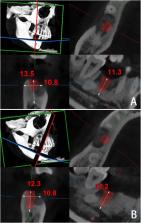
          CBCT images of 122 patients with implant in mandibular or maxillary first molar region were retrospectively collected. Two methods were used to measure sagittal height (SH), coronal height (CH), sagittal width (SW), and coronal width (CW) of implants. Method 1 (general method): the images were analyzed using the built-in software NNT 9.0 software. SHl, CHl, SWl, and CWl were measured on the reconstructed sagittal and coronal based on the radiologist’s own experience. Method 2 (triaxial rotation method): the raw data were demonstrated in Expert mode of NNT 9.0 software, in which the coronal axis and sagittal axis were rotated paralleling to the long axis of the implant for reconstruction, and then SH2, CH2, SW2, and CW2 were measured on the reconstructed sagittal and coronal images. The results of two methods were compared with the actual implant size (H0, W0). Paired T-test was performed for statistical analysis. Dahlberg formula was used to check the measurement error.

          Results

          For method 1, there was no significant differences between SHl and H0 (P > 0.05), but significant differences between CHl and H0, SWl and W0, and CWl and W0 (P < 0.05). For method 2, there were no significant differences between all measurements and actual size (P > 0.05). The random error range measured using Dahlberg formula was 0.157–1.171 mm for general method and 0.017–0.05 mm for triaxial rotation method.

          Conclusion

          The triaxial rotation method is accurate for implant height and width measurements on CBCT images and could be used in pre-operatively bone height and width measurement of potential implant sites.

          Related collections

          Most cited references23

          • Record: found
          • Abstract: found
          • Article: not found

          Effectiveness of Contour Augmentation with Guided Bone Regeneration: 10-Year Results

          In aesthetic sites, the integrity of the facial bone wall dimension in the anterior maxilla is jeopardized by physiologic and structural changes postextraction. An effective regenerative protocol is key to reestablish and maintain the hard and soft tissue dimensions over time. The present prospective case series study examined the effectiveness of early implant placement with simultaneous contour augmentation through guided bone regeneration with a 2-layer composite graft in postextraction single-tooth sites over an observation period of 10 y among 20 patients. The median peri-implant bone loss was 0.35 mm between the 1- and 10-y examination. A success rate of 95% was obtained, with pleasing aesthetic outcomes and a high median Pink Esthetic Score (8). Implant crowns (ICs) revealed significant median facial recession between IC10y and IC1y (0.17 mm). The facial bone wall dimensions were assessed by preoperative cone beam computed tomography and 2 subsequent scans taken at 6 and 10 y. The median facial bone wall thickness increased significantly from 0 mm at surgery to 1.67 mm at the 10-y examination. The facial vertical bone wall peak (DIC) was located at a median distance of 0.16 mm coronal to the implant shoulder. The facial vertical bone loss of DIC amounted to 0.02 mm between 6 and 10 y. Equivalence testing was performed for the null hypothesis of a difference of >0.2 mm per year between 2 respective time points, showing stable bone conditions. Modulating factors influencing the regenerative outcomes at 10 y were the preoperative proximal crest width and soft tissue thickness. In conclusion, the present study confirmed the long-term effectiveness of early implant placement with simultaneous contour augmentation through guided bone regeneration with a 2-layer composite graft in postextraction single-tooth sites offering stable bone conditions with low risks of mucosal recessions over an observation period of 10 y ( ClinicalTrials.gov NCT03252106).
            Bookmark
            • Record: found
            • Abstract: found
            • Article: not found

            Use of cone beam computed tomography in implant dentistry: the International Congress of Oral Implantologists consensus report.

            The International Congress of Oral Implantologists has supported the development of this consensus report involving the use of Cone Beam Computed Tomography (CBCT) in implant dentistry with the intent of providing scientifically based guidance to clinicians regarding its use as an adjunct to traditional imaging modalities. The literature regarding CBCT and implant dentistry was systematically reviewed. A PubMed search that included studies published between January 1, 2000, and July 31, 2011, was conducted. Oral presentations, in conjunction with these studies, were given by Dr. Erika Benavides, Dr. Scott Ganz, Dr. James Mah, Dr. Myung-Jin Kim, and Dr. David Hatcher at a meeting of the International Congress of Oral Implantologists in Seoul, Korea, on October 6-8, 2011. The studies published could be divided into four main groups: diagnostics, implant planning, surgical guidance, and postimplant evaluation. The literature supports the use of CBCT in dental implant treatment planning particularly in regards to linear measurements, three-dimensional evaluation of alveolar ridge topography, proximity to vital anatomical structures, and fabrication of surgical guides. Areas such as CBCT-derived bone density measurements, CBCT-aided surgical navigation, and postimplant CBCT artifacts need further research. ICOI RECOMMENDATIONS: All CBCT examinations, as all other radiographic examinations, must be justified on an individualized needs basis. The benefits to the patient for each CBCT scan must outweigh the potential risks. CBCT scans should not be taken without initially obtaining thorough medical and dental histories and performing a comprehensive clinical examination. CBCT should be considered as an imaging alternative in cases where the projected implant receptor or bone augmentation site(s) are suspect, and conventional radiography may not be able to assess the true regional three-dimensional anatomical presentation. The smallest possible field of view should be used, and the entire image volume should be interpreted.
              Bookmark
              • Record: found
              • Abstract: found
              • Article: not found

              Role of implant diameter on long-term survival of dental implants placed in posterior maxilla: a systematic review.

              We speculated that the long-term survival of narrow or conventional diameter (<5 mm) implants is higher than that of wide-diameter implants (≥ 5 mm) when placed in posterior atrophic maxillae. The aim of this paper was to systematically review indexed literature regarding the influence of implant diameter on long-term survival of dental implants placed in posterior maxilla.
                Bookmark

                Author and article information

                Contributors
                Journal
                Heliyon
                Heliyon
                Heliyon
                Elsevier
                2405-8440
                31 May 2024
                15 June 2024
                31 May 2024
                : 10
                : 11
                : e32076
                Affiliations
                [a ]Department of Dentomaxillofacial Radiology, Nanjing Stomatological Hospital, Affiliated Hospital of Medical School, Institute of Stomatology, Nanjing University, Nanjing, China
                [b ]Department of Stomatology, Shenzhen Longhua District Central Hospital, Shenzhen, Guangdong Province, China
                Author notes
                [* ]Corresponding author. Department of Dentomaxillofacial Radiology, Nanjing Stomatological Hospital, Affiliated Hospital of Medical School, Institute of Stomatology, Nanjing University, Zhong Yang Road 30, Nanjing City, Jiangsu Province, 210008, China. linzitong_710@ 123456163.com
                [1]

                Co-first author.

                Article
                S2405-8440(24)08107-6 e32076
                10.1016/j.heliyon.2024.e32076
                11168391
                38868001
                636d305d-ea23-4776-8ab6-f11949d8b539
                © 2024 The Authors. Published by Elsevier Ltd.

                This is an open access article under the CC BY-NC license (http://creativecommons.org/licenses/by-nc/4.0/).

                History
                : 6 February 2024
                : 11 May 2024
                : 28 May 2024
                Categories
                Research Article

                cone beam ct,sagittal,coronal,height,width,triaxial rotation method

                Comments

                Comment on this article