8
views
0
recommends
+1 Recommend
0 collections
    0
    shares
      • Record: found
      • Abstract: found
      • Article: found
      Is Open Access

      Cystatin C deficiency suppresses tumor growth in a breast cancer model through decreased proliferation of tumor cells

      research-article

      Read this article at

      Bookmark
          There is no author summary for this article yet. Authors can add summaries to their articles on ScienceOpen to make them more accessible to a non-specialist audience.

          Abstract

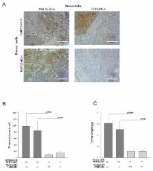
          Cysteine cathepsins are proteases that, in addition to their important physiological functions, have been associated with multiple pathologies, including cancer. Cystatin C (CstC) is a major endogenous inhibitor that regulates the extracellular activity of cysteine cathepsins. We investigated the role of cystatin C in mammary cancer using CstC knockout mice and a mouse model of breast cancer induced by expression of the polyoma middle T oncoprotein (PyMT) in the mammary epithelium. We showed that the ablation of CstC reduced the rate of mammary tumor growth. Notably, a decrease in the proliferation of CstC knockout PyMT tumor cells was demonstrated ex vivo and in vitro, indicating a role for this protease inhibitor in signaling pathways that control cell proliferation. An increase in phosphorylated p-38 was observed in CstC knockout tumors, suggesting a novel function for cystatin C in cancer development, independent of the TGF-β pathway. Moreover, proteomic analysis of the CstC wild-type and knockout PyMT primary cell secretomes revealed a decrease in the levels of 14-3-3 proteins in the secretome of knock-out cells, suggesting a novel link between cysteine cathepsins, cystatin C and 14-3-3 proteins in tumorigenesis, calling for further investigations.

          Related collections

          Most cited references69

          • Record: found
          • Abstract: found
          • Article: not found

          Immunological hallmarks of stromal cells in the tumour microenvironment.

          A dynamic and mutualistic interaction between tumour cells and the surrounding stroma promotes the initiation, progression, metastasis and chemoresistance of solid tumours. Far less understood is the relationship between the stroma and tumour-infiltrating leukocytes; however, emerging evidence suggests that the stromal compartment can shape antitumour immunity and responsiveness to immunotherapy. Thus, there is growing interest in elucidating the immunomodulatory roles of the stroma that evolve within the tumour microenvironment. In this Review, we discuss the evidence that stromal determinants interact with leukocytes and influence antitumour immunity, with emphasis on the immunological attributes of stromal cells that may foster their protumorigenic function.
            Bookmark
            • Record: found
            • Abstract: found
            • Article: not found

            Comparison of label-free methods for quantifying human proteins by shotgun proteomics.

            Measurements of mass spectral peak intensities and spectral counts are promising methods for quantifying protein abundance changes in shotgun proteomic analyses. We describe Serac, software developed to evaluate the ability of each method to quantify relative changes in protein abundance. Dynamic range and linearity using a three-dimensional ion trap were tested using standard proteins spiked into a complex sample. Linearity and good agreement between observed versus expected protein ratios were obtained after normalization and background subtraction of peak area intensity measurements and correction of spectral counts to eliminate discontinuity in ratio estimates. Peak intensity values useful for protein quantitation ranged from 10(7) to 10(11) counts with no obvious saturation effect, and proteins in replicate samples showed variations of less than 2-fold within the 95% range (+/-2sigma) when >or=3 peptides/protein were shared between samples. Protein ratios were determined with high confidence from spectral counts when maximum spectral counts were >or=4 spectra/protein, and replicates showed equivalent measurements well within 95% confidence limits. In further tests, complex samples were separated by gel exclusion chromatography, quantifying changes in protein abundance between different fractions. Linear behavior of peak area intensity measurements was obtained for peptides from proteins in different fractions. Protein ratios determined by spectral counting agreed well with those determined from peak area intensity measurements, and both agreed with independent measurements based on gel staining intensities. Overall spectral counting proved to be a more sensitive method for detecting proteins that undergo changes in abundance, whereas peak area intensity measurements yielded more accurate estimates of protein ratios. Finally these methods were used to analyze differential changes in protein expression in human erythroleukemia K562 cells stimulated under conditions that promote cell differentiation by mitogen-activated protein kinase pathway activation. Protein changes identified with p<0.1 showed good correlations with parallel measurements of changes in mRNA expression.
              Bookmark
              • Record: found
              • Abstract: found
              • Article: not found

              Perivascular M2 Macrophages Stimulate Tumor Relapse after Chemotherapy.

              Tumor relapse after chemotherapy-induced regression is a major clinical problem, because it often involves inoperable metastatic disease. Tumor-associated macrophages (TAM) are known to limit the cytotoxic effects of chemotherapy in preclinical models of cancer. Here, we report that an alternatively activated (M2) subpopulation of TAMs (MRC1(+)TIE2(Hi)CXCR4(Hi)) accumulate around blood vessels in tumors after chemotherapy, where they promote tumor revascularization and relapse, in part, via VEGF-A release. A similar perivascular, M2-related TAM subset was present in human breast carcinomas and bone metastases after chemotherapy. Although a small proportion of M2 TAMs were also present in hypoxic tumor areas, when we genetically ablated their ability to respond to hypoxia via hypoxia-inducible factors 1 and 2, tumor relapse was unaffected. TAMs were the predominant cells expressing immunoreactive CXCR4 in chemotherapy-treated mouse tumors, with the highest levels expressed by MRC1(+) TAMs clustering around the tumor vasculature. Furthermore, the primary CXCR4 ligand, CXCL12, was upregulated in these perivascular sites after chemotherapy, where it was selectively chemotactic for MRC1(+) TAMs. Interestingly, HMOX-1, a marker of oxidative stress, was also upregulated in perivascular areas after chemotherapy. This enzyme generates carbon monoxide from the breakdown of heme, a gas known to upregulate CXCL12. Finally, pharmacologic blockade of CXCR4 selectively reduced M2-related TAMs after chemotherapy, especially those in direct contact with blood vessels, thereby reducing tumor revascularization and regrowth. Our studies rationalize a strategy to leverage chemotherapeutic efficacy by selectively targeting this perivascular, relapse-promoting M2-related TAM cell population.
                Bookmark

                Author and article information

                Journal
                Oncotarget
                Oncotarget
                Oncotarget
                ImpactJ
                Oncotarget
                Impact Journals LLC
                1949-2553
                26 September 2017
                24 April 2017
                : 8
                : 43
                : 73793-73809
                Affiliations
                1 Department of Biochemistry and Molecular and Structural Biology, Jozef Stefan Institute, SI-1000 Ljubljana, Slovenia
                2 Jozef Stefan International Postgraduate School, Sl-1000 Ljubljana, Slovenia
                3 Department of Clinical Chemistry, Laboratory Medicine, University Hospital, SE-22185 Lund, Sweden
                4 Centre of Excellence for Integrated Approaches in Chemistry and Biology of Proteins, SI-1000 Ljubljana, Slovenia
                5 Faculty of Chemistry and Chemical Technology, University of Ljubljana, SI-1000 Ljubljana, Slovenia
                6 Current address: CytomX Therapeutics, Inc., South San Francisco, CA 94080, USA
                Author notes
                Correspondence to: Olga Vasiljeva, olga.vasiljeva@ 123456gmail.com
                Article
                17379
                10.18632/oncotarget.17379
                5650301
                29088746
                62853c3f-3126-4312-91f5-9191bdc63fb1
                Copyright: © 2017 Završnik et al.

                This is an open-access article distributed under the terms of the Creative Commons Attribution License 3.0 (CC BY 3.0), which permits unrestricted use, distribution, and reproduction in any medium, provided the original author and source are credited.

                History
                : 9 December 2016
                : 22 March 2017
                Categories
                Research Paper

                Oncology & Radiotherapy
                cathepsin inhibitor,cystatin c,breast cancer,mouse model
                Oncology & Radiotherapy
                cathepsin inhibitor, cystatin c, breast cancer, mouse model

                Comments

                Comment on this article