9
views
0
recommends
+1 Recommend
0 collections
    0
    shares
      • Record: found
      • Abstract: found
      • Article: found
      Is Open Access

      Establishment of Novel Limbus-Derived, Highly Proliferative ABCG2 +/ABCB5 + Limbal Epithelial Stem Cell Cultures

      research-article
      1 , 2 , 1 , 1 , 1 , 3 ,
      Stem Cells International
      Hindawi

      Read this article at

      Bookmark
          There is no author summary for this article yet. Authors can add summaries to their articles on ScienceOpen to make them more accessible to a non-specialist audience.

          Abstract

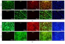
          Homeostasis and regeneration of corneal epithelia are sustained by limbal epithelial stem cells (LESCs); thus, an LESC deficiency is a major cause of blindness worldwide. Despite the generally promising results of cultivated LESC transplantation, it has been limited by variations in long-term success rates, the use of xenogeneic and undefined culture components, and a scarcity of donor tissues. In this study, we identified the culture conditions required to expand LESCs in vitro and established human limbus-derived highly proliferative ABCG2 +/ABCB5 + double-positive LESCs. These LESCs exhibited the LESC marker profile and differentiated into corneal epithelial cells. In addition, cultured LESCs expressed high levels of the stem cell markers Sox2, Oct4, c-Myc, and Klf4, had high telomerase activity, and had stable, normal genomes. These results suggest that our novel cultivation protocol affects the phenotype and differentiation capacity of LESCs. From the limbus, which contains a heterogenous cell population, we have derived highly proliferative ABCG2 +/ABCB5 + double-positive cells with the ability to differentiate into corneal epithelial cells. This study opens a new avenue for investigation of the molecular mechanism of LESC maintenance and expansion in vitro and may impact the treatment of corneal disease, particularly corneal blindness due to an LESC deficiency.

          Related collections

          Most cited references45

          • Record: found
          • Abstract: found
          • Article: not found

          In vitro reprogramming of fibroblasts into a pluripotent ES-cell-like state.

          Nuclear transplantation can reprogramme a somatic genome back into an embryonic epigenetic state, and the reprogrammed nucleus can create a cloned animal or produce pluripotent embryonic stem cells. One potential use of the nuclear cloning approach is the derivation of 'customized' embryonic stem (ES) cells for patient-specific cell treatment, but technical and ethical considerations impede the therapeutic application of this technology. Reprogramming of fibroblasts to a pluripotent state can be induced in vitro through ectopic expression of the four transcription factors Oct4 (also called Oct3/4 or Pou5f1), Sox2, c-Myc and Klf4. Here we show that DNA methylation, gene expression and chromatin state of such induced reprogrammed stem cells are similar to those of ES cells. Notably, the cells-derived from mouse fibroblasts-can form viable chimaeras, can contribute to the germ line and can generate live late-term embryos when injected into tetraploid blastocysts. Our results show that the biological potency and epigenetic state of in-vitro-reprogrammed induced pluripotent stem cells are indistinguishable from those of ES cells.
            Bookmark
            • Record: found
            • Abstract: found
            • Article: not found

            Serial cultivation of strains of human epidermal keratinocytes: the formation of keratinizing colonies from single cells.

            Human diploid epidermis epidermal cells have been successfully grown in serial culture. To initiate colony formation, they require the presence of fibroblasts, but proliferation of fibroblasts must be controlled so that the epidermal cell population is not overgrown. Both conditions can be achieved by the use of lethally irradiated 3T3 cells at the correct density. When trypsinized human skin cells are plated together with the 3T3 cells, the growth of the human fibroblasts is largely suppressed, but epidermal cells grow from single cells into colonies. Each colony consists of keratinocytes ultimately forming a stratified squamous epithelium in which the dividing cells are confined to the lowest layer(s). Hydrocortisone is added to the medium, since in secondary and subsequent subcultures it makes the colony morphology more oderly and distinctive, and maintains proliferation at a slightly greater rate. Under these culture conditions, it is possible to isolate keratinocyte clones free of viable fibroblasts. Like human diploid fibroblasts, human diploid keratinocytes appear to have a finite culture lifetime. For 7 strains studied, the culture lifetime ranged from 20-50 cell generations. The plating efficiency of the epidermal cells taken directly from skin was usually 0.1-1.0%. On subsequent transfer of the cultures initiated from newborns, the plating efficiency rose to 10% or higher, but was most often in the range of 1-5% and dropped sharply toward the end of their culture life. The plating efficiency and culture lifetime were lower for keratinocytes of older persons.
              Bookmark
              • Record: found
              • Abstract: found
              • Article: not found

              Three clonal types of keratinocyte with different capacities for multiplication.

              Colony-forming human epidermal cells are heterogeneous in their capacity for sustained growth. Once a clone has been derived from a single cell, its growth potential can be estimated from the colony types resulting from a single plating, and the clone can be assigned to one of three classes. The holoclone has the greatest reproductive capacity: under standard conditions, fewer than 5% of the colonies formed by the cells of a holoclone abort and terminally differentiate. The paraclone contains exclusively cells with a short replicative lifespan (not more than 15 cell generations), after which they uniformly abort and terminally differentiate. The third type of clone, the meroclone, contains a mixture of cells of different growth potential and is a transitional stage between the holoclone and the paraclone. The incidence of the different clonal types is affected by aging, since cells originating from the epidermis of older donors give rise to a lower proportion of holoclones and a higher proportion of paraclones.
                Bookmark

                Author and article information

                Contributors
                Journal
                Stem Cells Int
                Stem Cells Int
                SCI
                Stem Cells International
                Hindawi
                1687-966X
                1687-9678
                2017
                5 November 2017
                : 2017
                : 7678637
                Affiliations
                1Department of Ophthalmology, Corneal Dystrophy Research Institute, Yonsei University College of Medicine, Seoul, Republic of Korea
                2Institute of Vision Research, Severance Biomedical Science Institute, Yonsei University College of Medicine, Seoul, Republic of Korea
                3Department of Obstetrics and Gynecology, Institute of Women's Life Medical Science, Yonsei University College of Medicine, Seoul, Republic of Korea
                Author notes

                Academic Editor: Ming Li

                Author information
                http://orcid.org/0000-0002-1453-8042
                http://orcid.org/0000-0003-1694-8405
                Article
                10.1155/2017/7678637
                5694571
                623b33a9-a6bd-442f-9352-9578bde5f8d5
                Copyright © 2017 Eung Kweon Kim et al.

                This is an open access article distributed under the Creative Commons Attribution License, which permits unrestricted use, distribution, and reproduction in any medium, provided the original work is properly cited.

                History
                : 26 June 2017
                : 27 September 2017
                : 12 October 2017
                Funding
                Funded by: Ministry of Education
                Award ID: NRF-2016R1D1A1B03933337
                Funded by: National Research Foundation of Korea
                Categories
                Research Article

                Molecular medicine
                Molecular medicine

                Comments

                Comment on this article