10
views
0
recommends
+1 Recommend
0 collections
    0
    shares
      • Record: found
      • Abstract: found
      • Article: found
      Is Open Access

      Isolation and characterization of equine endometrial mesenchymal stromal cells

      research-article

      Read this article at

      Bookmark
          There is no author summary for this article yet. Authors can add summaries to their articles on ScienceOpen to make them more accessible to a non-specialist audience.

          Abstract

          Background

          Equine mesenchymal stromal/stem cells (MSCs) are most commonly harvested from bone marrow (BM) or adipose tissue, requiring the use of surgical procedures. By contrast, the uterus can be accessed nonsurgically, and may provide a more readily available cell source. While human endometrium is known to harbor mesenchymal precursor cells, MSCs have not been identified in equine endometrium. This study reports the isolation, culture, and characterization of MSCs from equine endometrium.

          Methods

          The presence of MSC and pericyte markers in endometrial sections was determined using immunohistochemistry. Stromal cells were harvested and cultured after separation of epithelial cells from endometrial fragments using Mucin-1-bound beads. For comparison, MSCs were also harvested from BM. The expression of surface markers in endometrial and BM-derived MSCs was characterized using flow cytometry and quantitative polymerase chain reaction. MSCs were differentiated in vitro into adipogenic, chondrogenic, osteogenic, and smooth muscle lineages.

          Results

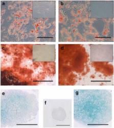
          Typical markers of MSCs (CD29, CD44, CD90, and CD105) and pericytes (NG2 and CD146) were localized in the equine endometrium. Both endometrial and BM MSCs grew clonally and robustly expressed MSC and pericyte markers in culture while showing greatly reduced or negligible expression of hematopoietic markers (CD45, CD34) and MHC-II. Additionally, both endometrial and BM MSCs differentiated into adipogenic, osteogenic, and chondrogenic lineages in vitro, and endometrial MSCs had a distinct ability to undergo smooth muscle differentiation.

          Conclusions

          We have demonstrated for the first time the presence of cells in equine endometrium that fulfill the definition of MSCs. The equine endometrium may provide an alternative, easily accessible source of MSCs, not only for therapeutic regeneration of the uterus, but also for other tissues where MSCs from other sources are currently being used therapeutically.

          Related collections

          Most cited references35

          • Record: found
          • Abstract: found
          • Article: not found

          Mesenchymal progenitor cells in human umbilical cord blood.

          Haemopoiesis is sustained by two main cellular components, the haematopoietic cells (HSCs) and the mesenchymal progenitor cells (MPCs). MPCs are multipotent and are the precursors for marrow stroma, bone, cartilage, muscle and connective tissues. Although the presence of HSCs in umbilical cord blood (UCB) is well known, that of MPCs has been not fully evaluated. In this study, we examined the ability of UCB harvests to generate in culture cells with characteristics of MPCs. Results showed that UCB-derived mononuclear cells, when set in culture, gave rise to adherent cells, which exhibited either an osteoclast- or a mesenchymal-like phenotype. Cells with the osteoclast phenotype were multinucleated, expressed TRAP activity and antigens CD45 and CD51/CD61. In turn, cells with the mesenchymal phenotype displayed a fibroblast-like morphology and expressed several MPC-related antigens (SH2, SH3, SH4, ASMA, MAB 1470, CD13, CD29 and CD49e). Our results suggest that preterm, as compared with term, cord blood is richer in mesenchymal progenitors, similar to haematopoietic progenitors.
            Bookmark
            • Record: found
            • Abstract: found
            • Article: found
            Is Open Access

            No Identical “Mesenchymal Stem Cells” at Different Times and Sites: Human Committed Progenitors of Distinct Origin and Differentiation Potential Are Incorporated as Adventitial Cells in Microvessels

            Summary A widely shared view reads that mesenchymal stem/stromal cells (“MSCs”) are ubiquitous in human connective tissues, can be defined by a common in vitro phenotype, share a skeletogenic potential as assessed by in vitro differentiation assays, and coincide with ubiquitous pericytes. Using stringent in vivo differentiation assays and transcriptome analysis, we show that human cell populations from different anatomical sources, regarded as “MSCs” based on these criteria and assumptions, actually differ widely in their transcriptomic signature and in vivo differentiation potential. In contrast, they share the capacity to guide the assembly of functional microvessels in vivo, regardless of their anatomical source, or in situ identity as perivascular or circulating cells. This analysis reveals that muscle pericytes, which are not spontaneously osteochondrogenic as previously claimed, may indeed coincide with an ectopic perivascular subset of committed myogenic cells similar to satellite cells. Cord blood-derived stromal cells, on the other hand, display the unique capacity to form cartilage in vivo spontaneously, in addition to an assayable osteogenic capacity. These data suggest the need to revise current misconceptions on the origin and function of so-called “MSCs,” with important applicative implications. The data also support the view that rather than a uniform class of “MSCs,” different mesoderm derivatives include distinct classes of tissue-specific committed progenitors, possibly of different developmental origin.
              Bookmark
              • Record: found
              • Abstract: found
              • Article: not found

              Clonogenicity of human endometrial epithelial and stromal cells.

              The human endometrium regenerates from the lower basalis layer, a germinal compartment that persists after menstruation to give rise to the new upper functionalis layer. Because adult stem cells are present in tissues that undergo regeneration, we hypothesized that human endometrium contains small populations of epithelial and stromal stem cells responsible for cyclical regeneration of endometrial glands and stroma and that these cells would exhibit clonogenicity, a stem-cell property. The aims of this study were to determine 1) the clonogenic activity of human endometrial epithelial and stromal cells, 2) which growth factors support this clonogenic activity, and 3) determine the cellular phenotypes of the clones. Endometrial tissue was obtained from women undergoing hysterectomy. Purified single- cell suspensions of epithelial and stromal cells were cultured at cloning density (300-500/cm(2)) in serum medium or in serum- free medium supplemented with one of eight growth factors. Small numbers of epithelial (0.22%) and stromal cells (1.25%) initiated colonies in serum-containing medium. The majority of colonies were small, containing large, loosely arranged cells, and 37% of epithelial and 1 in 60 of stromal colonies were classified as large, comprising small, densely packed cells. In serum-free medium, transforming growth factor-alpha (TGF alpha), epidermal growth factor (EGF), platelet-derived growth factor-BB (PDGF-BB) strongly supported clonogenicity of epithelial cells, while leukemia-inhibitory factor (LIF), hepatocyte growth factor (HGF), stem-cell factor (SCF), insulin-like growth factor-I (IGF- I) were weakly supportive, and basic fibroblast growth factor (bFGF) was without effect. TGF alpha, EGF, PDGF-BB, and bFGF supported stromal cell clonogenicity, while HGF, SCF, LIF, and IGF- I were without effect. Small epithelial colonies expressed three epithelial markers but not stromal markers; however, large epithelial colonies showed little reactivity for all markers except alpha(6)-integrin. All stromal colonies contained fibroblasts, expressing stromal markers, and in some colonies, myofibroblasts were also identified. This analysis of human endometrium has demonstrated the presence of rare clonogenic epithelial and stromal cells with high proliferative potential, providing the first evidence for the existence of putative endometrial epithelial and stromal stem cells.
                Bookmark

                Author and article information

                Contributors
                Elisabeth.Rink@vetmeduni.ac.at
                Karin.Amilon@roslin.ed.ac.uk
                Cristina.Esteves@roslin.ed.ac.uk
                HFrench@RossU.edu
                elaine.watson111@btinternet.com
                Christine.Aurich@vetmeduni.ac.at
                +44 (0)131-6519161 , Xavier.Donadeu@roslin.ed.ac.uk
                Journal
                Stem Cell Res Ther
                Stem Cell Res Ther
                Stem Cell Research & Therapy
                BioMed Central (London )
                1757-6512
                12 July 2017
                12 July 2017
                2017
                : 8
                : 166
                Affiliations
                [1 ]ISNI 0000 0004 1776 0209, GRID grid.412247.6, , Ross University School of Veterinary Medicine, ; Basseterre, Saint Kitts and Nevis
                [2 ]ISNI 0000 0004 1936 7988, GRID grid.4305.2, The Roslin Institute, , University of Edinburgh, ; Edinburgh, EH25 9RG UK
                [3 ]ISNI 0000 0000 9686 6466, GRID grid.6583.8, , University of Veterinary Medicine, ; 1220 Vienna, Austria
                [4 ]ISNI 0000 0004 1936 7988, GRID grid.4305.2, The Roslin Institute, , University of Edinburgh, ; Easter Bush, Midlothian, EH25 9RG UK
                Article
                616
                10.1186/s13287-017-0616-0
                5506676
                28701175
                60546ebf-b3c1-4bfc-9f64-22c36317e270
                © The Author(s). 2017

                Open AccessThis article is distributed under the terms of the Creative Commons Attribution 4.0 International License ( http://creativecommons.org/licenses/by/4.0/), which permits unrestricted use, distribution, and reproduction in any medium, provided you give appropriate credit to the original author(s) and the source, provide a link to the Creative Commons license, and indicate if changes were made. The Creative Commons Public Domain Dedication waiver ( http://creativecommons.org/publicdomain/zero/1.0/) applies to the data made available in this article, unless otherwise stated.

                History
                : 1 February 2017
                : 14 June 2017
                : 20 June 2017
                Funding
                Funded by: Ross University School of Veterinary Medicine
                Funded by: FundRef http://dx.doi.org/10.13039/501100000268, Biotechnology and Biological Sciences Research Council;
                Funded by: FundRef http://dx.doi.org/10.13039/501100001280, Horserace Betting Levy Board;
                Award ID: Prj768
                Categories
                Research
                Custom metadata
                © The Author(s) 2017

                Molecular medicine
                mesenchymal stem cells,endometrium,equine,horse
                Molecular medicine
                mesenchymal stem cells, endometrium, equine, horse

                Comments

                Comment on this article