1
views
0
recommends
+1 Recommend
0 collections
    0
    shares
      • Record: found
      • Abstract: found
      • Article: found
      Is Open Access

      Caspase/AIF/apoptosis pathway: a new target of puerarin for diabetes mellitus therapy

      research-article

      Read this article at

      Bookmark
          There is no author summary for this article yet. Authors can add summaries to their articles on ScienceOpen to make them more accessible to a non-specialist audience.

          Abstract

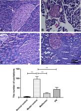
          Pancreatic β cell damage is one of the crucial factors responsible for the development of type 2 diabetes mellitus (T2DM). Previous studies have suggested that puerarin (PR) could regulate the activities of the mitochondrial respiratory chain complex in diabetic nephropathy (DN); however, whether PR can inhibit pancreatic β-cell apoptosis in T2DM remains to be elucidated. In the present study, T2DM mice induced by high-fat diet and streptozotocin (STZ) injection were used as a working model to investigate the mechanism of PR on pancreatic β cell apoptosis. The results showed that PR decreased the serum fasting blood glucose (FBG), total cholesterol (TC), triglyceride (TG) and low-density lipoprotein (LDL) levels but significantly increased the fasting blood insulin (FINS) and high-density lipoprotein (HDL) levels. Furthermore, decreased caspase-3, 8, 9 and apoptosis-inducing factor (AIF) proteins in the pancreas were detected by Western blot analysis. Terminal deoxynucleotidyl transferase-mediated dUTP nick end labelling (TUNEL) staining demonstrated that the pancreatic β cell apoptosis was inhibited by PR. Furthermore, PR improved the histopathological changes in pancreatic tissue in T2DM mice. Collectively, the data show that PR can protect the β cells from apoptotic death in a mouse model of T2DM through regulating the expression of apoptosis-related protein-AIF and caspase family proteins.

          Related collections

          Most cited references32

          • Record: found
          • Abstract: found
          • Article: not found

          Molecular characterization of mitochondrial apoptosis-inducing factor.

          Mitochondria play a key part in the regulation of apoptosis (cell death). Their intermembrane space contains several proteins that are liberated through the outer membrane in order to participate in the degradation phase of apoptosis. Here we report the identification and cloning of an apoptosis-inducing factor, AIF, which is sufficient to induce apoptosis of isolated nuclei. AIF is a flavoprotein of relative molecular mass 57,000 which shares homology with the bacterial oxidoreductases; it is normally confined to mitochondria but translocates to the nucleus when apoptosis is induced. Recombinant AIF causes chromatin condensation in isolated nuclei and large-scale fragmentation of DNA. It induces purified mitochondria to release the apoptogenic proteins cytochrome c and caspase-9. Microinjection of AIF into the cytoplasm of intact cells induces condensation of chromatin, dissipation of the mitochondrial transmembrane potential, and exposure of phosphatidylserine in the plasma membrane. None of these effects is prevented by the wide-ranging caspase inhibitor known as Z-VAD.fmk. Overexpression of Bcl-2, which controls the opening of mitochondrial permeability transition pores, prevents the release of AIF from the mitochondrion but does not affect its apoptogenic activity. These results indicate that AIF is a mitochondrial effector of apoptotic cell death.
            Bookmark
            • Record: found
            • Abstract: found
            • Article: not found

            Pancreatic β cell dedifferentiation as a mechanism of diabetic β cell failure.

            Diabetes is associated with β cell failure. But it remains unclear whether the latter results from reduced β cell number or function. FoxO1 integrates β cell proliferation with adaptive β cell function. We interrogated the contribution of these two processes to β cell dysfunction, using mice lacking FoxO1 in β cells. FoxO1 ablation caused hyperglycemia with reduced β cell mass following physiologic stress, such as multiparity and aging. Surprisingly, lineage-tracing experiments demonstrated that loss of β cell mass was due to β cell dedifferentiation, not death. Dedifferentiated β cells reverted to progenitor-like cells expressing Neurogenin3, Oct4, Nanog, and L-Myc. A subset of FoxO1-deficient β cells adopted the α cell fate, resulting in hyperglucagonemia. Strikingly, we identify the same sequence of events as a feature of different models of murine diabetes. We propose that dedifferentiation trumps endocrine cell death in the natural history of β cell failure and suggest that treatment of β cell dysfunction should restore differentiation, rather than promoting β cell replication. Copyright © 2012 Elsevier Inc. All rights reserved.
              Bookmark
              • Record: found
              • Abstract: found
              • Article: not found

              Essential role of the mitochondrial apoptosis-inducing factor in programmed cell death.

              Programmed cell death is a fundamental requirement for embryogenesis, organ metamorphosis and tissue homeostasis. In mammals, release of mitochondrial cytochrome c leads to the cytosolic assembly of the apoptosome-a caspase activation complex involving Apaf1 and caspase-9 that induces hallmarks of apoptosis. There are, however, mitochondrially regulated cell death pathways that are independent of Apaf1/caspase-9. We have previously cloned a molecule associated with programmed cell death called apoptosis-inducing factor (AIF). Like cytochrome c, AIF is localized to mitochondria and released in response to death stimuli. Here we show that genetic inactivation of AIF renders embryonic stem cells resistant to cell death after serum deprivation. Moreover, AIF is essential for programmed cell death during cavitation of embryoid bodies-the very first wave of cell death indispensable for mouse morphogenesis. AIF-dependent cell death displays structural features of apoptosis, and can be genetically uncoupled from Apaf1 and caspase-9 expression. Our data provide genetic evidence for a caspase-independent pathway of programmed cell death that controls early morphogenesis.
                Bookmark

                Author and article information

                Contributors
                kqliangtao@126.com
                Journal
                Mol Biol Rep
                Mol Biol Rep
                Molecular Biology Reports
                Springer Netherlands (Dordrecht )
                0301-4851
                1573-4978
                21 June 2019
                21 June 2019
                2019
                : 46
                : 5
                : 4787-4797
                Affiliations
                [1 ]GRID grid.256607.0, ISNI 0000 0004 1798 2653, College of Stomatology of Guangxi Medical University, ; No. 10, Shuangyong Road, Nanning, 530021 People’s Republic of China
                [2 ]GRID grid.413431.0, Affiliated Tumor Hospital of Guangxi Medical University, , Institute of Cancer Prevention and Treatment of Guangxi Zhuang Autonomous Region, ; Nanning, 530021 Guangxi People’s Republic of China
                [3 ]Department of Clinical Pharmacy, Hospital of Guangxi Zhuang Autonomous Region, Nanning, 530021 Guangxi People’s Republic of China
                [4 ]GRID grid.256607.0, ISNI 0000 0004 1798 2653, Guangxi Medical University, ; Nanning, 530021 Guangxi People’s Republic of China
                Article
                4925
                10.1007/s11033-019-04925-1
                8782775
                31228042
                602f3a00-5595-45ad-b465-1f896be16f3f
                © The Author(s) 2019

                Open AccessThis article is distributed under the terms of the Creative Commons Attribution 4.0 International License ( http://creativecommons.org/licenses/by/4.0/), which permits unrestricted use, distribution, and reproduction in any medium, provided you give appropriate credit to the original author(s) and the source, provide a link to the Creative Commons license, and indicate if changes were made.

                History
                : 9 April 2019
                : 14 June 2019
                Funding
                Funded by: Guangxi Natural Science Foundation
                Award ID: 2018GXNSFBA281037
                Award ID: 2018GXNSFBA281096
                Award Recipient :
                Funded by: FundRef http://dx.doi.org/10.13039/501100012543, Youth Science Foundation of Guangxi Medical University;
                Award ID: GXMUYSF201635
                Award Recipient :
                Funded by: Guangxi Middle School and Young Teachers' Basic Ability Improvement Project
                Award ID: 2018KY0128
                Award Recipient :
                Categories
                Original Article
                Custom metadata
                © Springer Nature B.V. 2019

                Molecular biology
                puerarin,type 2 diabetes mellitus,apoptosis-inducing factor,caspase
                Molecular biology
                puerarin, type 2 diabetes mellitus, apoptosis-inducing factor, caspase

                Comments

                Comment on this article