20
views
0
recommends
+1 Recommend
0 collections
    0
    shares
      • Record: found
      • Abstract: found
      • Article: found
      Is Open Access

      Nematode infection in liver of the fish Gymnotus inaequilabiatus (Gymnotiformes: Gymnotidae) from the Pantanal Region in Brazil: pathobiology and inflammatory response

      research-article

      Read this article at

      Bookmark
          There is no author summary for this article yet. Authors can add summaries to their articles on ScienceOpen to make them more accessible to a non-specialist audience.

          Abstract

          Background

          A survey on endoparasitic helminths from freshwater fishes in the Pantanal Region (Mato Grosso do Sul, Brazil) revealed the occurrence of third-larval stage of the nematode Brevimulticaecum sp. (Heterocheilidae) in most organs of Gymnotus inaequilabiatus (Gymnotidae) also known by the local name tuvira. The aim of the present study was to examine Brevimulticaecum sp.-infected tuvira liver at the ultrastructural level and clarify the nature of granulomas and the cellular elements involved in the immune response to nematode larvae.

          Methods

          Thirty-eight adult specimens of tuvira from Porto Morrinho, were acquired in January and March 2016. Infected and uninfected liver tissues were fixed and prepared for histological and ultrastructure investigations.

          Results

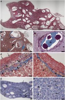
          The prevalence of infection of tuvira liver by the nematode larvae was 95 %, with an intensity of infection ranging from 4 to 343 larvae (mean ± SD: 55.31 ± 73.94 larvae per liver). In livers with high numbers of nematode larvae, almost entire hepatic tissue was occupied by the parasites. Hepatocytes showed slight to mild degenerative changes and accumulation of pigments. Parasite larvae were surrounded by round to oval granulomas, the result of focal host tissue response to the infection. Each granuloma was typically formed by three concentric layers: an outer layer of fibrous connective tissue with thin elongated fibroblasts; a middle layer of mast cells entrapped in a thin fibroblast-connective mesh; and an inner layer of densely packed epithelioid cells, displaying numerous desmosomes between each other. Numerous macrophage aggregates occurred in the granulomas and in the parenchyma.

          Conclusions

          Our results in tuvira showed that the larvae were efficiently sequestered within the granulomas, most of the inflammatory components were confined within the thickness of the granuloma, and the parenchyma was relatively free of immune cells and without fibrosis. Presumably this focal encapsulation of the parasites permits uninfected portions of liver to maintain its functions and allows the survival of the host.

          Related collections

          Most cited references69

          • Record: found
          • Abstract: found
          • Article: not found
          Is Open Access

          The zebrafish as a model to study intestinal inflammation.

          Starting out as a model for developmental biology, during the last decade, zebrafish have also gained the attention of the immunologists and oncologists. Due to its small size, high fecundity and full annotation of its genome, the zebrafish is an attractive model system. The fact that fish are transparent early in life combined with the growing list of immune cell reporter fish, enables in vivo tracking of immune responses in a complete organism. Since zebrafish develop ex utero from a fertilized egg, immune development can be monitored from the start of life. Given that several gut functions and immune genes are conserved between zebrafish and mammals, the zebrafish is an interesting model organism to investigate fundamental processes underlying intestinal inflammation and injury. This review will first provide some background on zebrafish intestinal development, bacterial colonization and immunity, showing the similarities and differences compared to mammals. This will be followed by an overview of the existing models for intestinal disease, and concluded by future perspectives in light of the newest technologies and insights.
            Bookmark
            • Record: found
            • Abstract: found
            • Article: not found

            Immune system and immune responses in fish and their role in comparative immunity study: a model for higher organisms.

            The basal position of fish in vertebrate phylogeny makes them very attractive for genomic and functional comparative immunity studies. Adaptive immunity arose early in vertebrate evolution, 450 million years ago between the divergence of cyclostomes and cartilaginous fish. The fundamental immune molecules, which include Ag-recognizing lymphocytes, immunoglobulins (Abs and Ig-family TCR), MHC products, and recombination-activating (RAG) 1 and 2 genes and the recombination mechanisms (cause of diversity in TCRs and Igs) are similar in fish and mammals. These molecules and their immune response mechanisms unravelled the primordial vertebrate immune system repertoire and adaptive radiations. Moreover, screening of animal models like zebrafish has a great importance to discover genes involved in T cell development, thymic organogenesis, and in immunity to infections. The zebrafish model may also be useful for cancer research due to its various features like rapid development, tractable genetics, ease in in vivo imaging and chemical screening. Copyright © 2012 Elsevier B.V. All rights reserved.
              Bookmark
              • Record: found
              • Abstract: found
              • Article: not found

              Innate host defense mechanisms of fish against viruses and bacteria.

              A Ellis (2001)
              The integumental defenses provide a physical and chemical barrier to the attachment and penetration of microbes. Besides the entrapping and sloughing of microbes in the mucus, the latter contains many antibacterial substances including anti-bacterial peptides, lysozyme, lectins and proteases. The gastro-intestinal tract is a hostile environment of acids, bile salts and enzymes able to inactivate and digest many viruses and bacteria. In most cases the integumental defenses are sufficient to protect against even quite virulent organisms which often only produce disease when the integument has been physically damaged. If a microbe gains access to the tissues of the fish, it is met with an array of soluble and cellular defenses. The complement system, present in the blood plasma, plays a central role in recognising bacteria and its activated products may lyse the bacterial cells, initiate inflammation, induce the influx of phagocytes and enhance their phagocytic activity. Complement can be activated directly by bacterial products and constituents and also indirectly by other factors, principally C-reactive protein and lectins, which can also bind to the bacterial surface. Plasma also contains a number of factors which inhibit bacterial growth(e.g. transferrin and anti-proteases) or which are bactericidal e.g. lysozyme. Following the infection of fish with virus pathogens, infected cells produce interferon. This induces antiviral defenses in neighbouring cells which are then protected from becoming infected. Anti-viral cytotoxic cells are able to lyse virally infected cells and thus reduce the rate of multiplication of virus within them. Innate defenses thus provide a pre-existing and fast-acting system of protection which is non-specific and relatively temperature-independent and thus has several advantages over the slow-acting and temperature-dependent specific immune responses.
                Bookmark

                Author and article information

                Contributors
                dzb@unife.it
                carlos.fernandes@ufms.br
                gmgalindo@ig.com.br
                ctg@unife.it
                mmanera@unite.it
                jadepasquale@morphogenyx.com
                massimo.lorenzoni@unipg.it
                sara.bertin@student.unife.it
                grilsu@unife.it
                Journal
                Parasit Vectors
                Parasit Vectors
                Parasites & Vectors
                BioMed Central (London )
                1756-3305
                30 August 2016
                30 August 2016
                2016
                : 9
                : 1
                : 473
                Affiliations
                [1 ]Department of Life Sciences and Biotechnology, University of Ferrara, Ferrara, Italy
                [2 ]Laboratory of Pathology, CCBS, Federal University of Mato Grosso do Sul, Campo Grande, Brazil
                [3 ]Department of Food Science, University of Teramo, St. Crispi 212, 64100 Teramo, Italy
                [4 ]Morphogenyx Inc, PO Box 717, 11731 East Northport, NY USA
                [5 ]Department of Cellular and Environmental Biology, University of Perugia, St. Elce di Sotto 5, 06123 Perugia, Italy
                Article
                1772
                10.1186/s13071-016-1772-2
                5006381
                27576434
                5fd92c0b-c4ba-4af6-a683-c874a1ed6fb7
                © The Author(s). 2016

                Open AccessThis article is distributed under the terms of the Creative Commons Attribution 4.0 International License ( http://creativecommons.org/licenses/by/4.0/), which permits unrestricted use, distribution, and reproduction in any medium, provided you give appropriate credit to the original author(s) and the source, provide a link to the Creative Commons license, and indicate if changes were made. The Creative Commons Public Domain Dedication waiver ( http://creativecommons.org/publicdomain/zero/1.0/) applies to the data made available in this article, unless otherwise stated.

                History
                : 30 June 2016
                : 23 August 2016
                Funding
                Funded by: FundRef http://dx.doi.org/10.13039/501100007109, Università degli Studi di Ferrara;
                Award ID: FAR2015
                Award Recipient :
                Categories
                Research
                Custom metadata
                © The Author(s) 2016

                Parasitology
                fish immune response,nematode larvae,hepatic granuloma,histopathology
                Parasitology
                fish immune response, nematode larvae, hepatic granuloma, histopathology

                Comments

                Comment on this article