22
views
0
recommends
+1 Recommend
0 collections
    0
    shares
      • Record: found
      • Abstract: found
      • Article: found
      Is Open Access

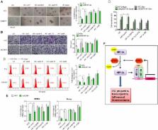
      SENP1 promotes hypoxia-induced cancer stemness by HIF-1α deSUMOylation and SENP1/HIF-1α positive feedback loop

      research-article

      Read this article at

      Bookmark
          There is no author summary for this article yet. Authors can add summaries to their articles on ScienceOpen to make them more accessible to a non-specialist audience.

          Abstract

          Objective

          We investigated the effect and mechanism of hypoxic microenvironment and hypoxia-inducible factors (HIFs) on hepatocellular carcinoma (HCC) cancer stemness.

          Design

          HCC cancer stemness was analysed by self-renewal ability, chemoresistance, expression of stemness-related genes and cancer stem cell (CSC) marker-positive cell population. Specific small ubiquitin-like modifier (SUMO) proteases 1 (SENP1) mRNA level was examined with quantitative PCR in human paired HCCs. Immunoprecipitation was used to examine the binding of proteins and chromatin immunoprecipitation assay to detect the binding of HIFs with hypoxia response element sequence. In vivo characterisation was performed in immunocompromised mice and stem cell frequency was analysed.

          Results

          We showed that hypoxia enhanced the stemness of HCC cells and hepatocarcinogenesis through enhancing HIF-1α deSUMOylation by SENP1 and increasing stabilisation and transcriptional activity of HIF-1α. Furthermore, we demonstrated that SENP1 is a direct target of HIF-1/2α and a previously unrecognised positive feedback loop exists between SENP1 and HIF-1α.

          Conclusions

          Taken together, our findings suggest the significance of this positive feedback loop between HIF-1α and SENP1 in contributing to the increased cancer stemness in HCC and hepatocarcinogenesis under hypoxia. Drugs that specifically target SENP1 may offer a potential novel therapeutic approach for HCC.

          Related collections

          Most cited references27

          • Record: found
          • Abstract: found
          • Article: not found

          Identification of pancreatic cancer stem cells.

          Emerging evidence has suggested that the capability of a tumor to grow and propagate is dependent on a small subset of cells within a tumor, termed cancer stem cells. Although data have been provided to support this theory in human blood, brain, and breast cancers, the identity of pancreatic cancer stem cells has not been determined. Using a xenograft model in which primary human pancreatic adenocarcinomas were grown in immunocompromised mice, we identified a highly tumorigenic subpopulation of pancreatic cancer cells expressing the cell surface markers CD44, CD24, and epithelial-specific antigen (ESA). Pancreatic cancer cells with the CD44(+)CD24(+)ESA(+) phenotype (0.2-0.8% of pancreatic cancer cells) had a 100-fold increased tumorigenic potential compared with nontumorigenic cancer cells, with 50% of animals injected with as few as 100 CD44(+)CD24(+)ESA(+) cells forming tumors that were histologically indistinguishable from the human tumors from which they originated. The enhanced ability of CD44(+)CD24(+)ESA(+) pancreatic cancer cells to form tumors was confirmed in an orthotopic pancreatic tail injection model. The CD44(+)CD24(+)ESA(+) pancreatic cancer cells showed the stem cell properties of self-renewal, the ability to produce differentiated progeny, and increased expression of the developmental signaling molecule sonic hedgehog. Identification of pancreatic cancer stem cells and further elucidation of the signaling pathways that regulate their growth and survival may provide novel therapeutic approaches to treat pancreatic cancer, which is notoriously resistant to standard chemotherapy and radiation.
            Bookmark
            • Record: found
            • Abstract: found
            • Article: not found

            Protein modification by SUMO.

            Small ubiquitin-related modifier (SUMO) family proteins function by becoming covalently attached to other proteins as post-translational modifications. SUMO modifies many proteins that participate in diverse cellular processes, including transcriptional regulation, nuclear transport, maintenance of genome integrity, and signal transduction. Reversible attachment of SUMO is controlled by an enzyme pathway that is analogous to the ubiquitin pathway. The functional consequences of SUMO attachment vary greatly from substrate to substrate, and in many cases are not understood at the molecular level. Frequently SUMO alters interactions of substrates with other proteins or with DNA, but SUMO can also act by blocking ubiquitin attachment sites. An unusual feature of SUMO modification is that, for most substrates, only a small fraction of the substrate is sumoylated at any given time. This review discusses our current understanding of how SUMO conjugation is controlled, as well as the roles of SUMO in a number of biological processes.
              Bookmark
              • Record: found
              • Abstract: found
              • Article: not found

              Single-cell cloning of colon cancer stem cells reveals a multi-lineage differentiation capacity.

              Colon carcinoma is one of the leading causes of death from cancer and is characterized by a heterogenic pool of cells with distinct differentiation patterns. Recently, it was reported that a population of undifferentiated cells from a primary tumor, so-called cancer stem cells (CSC), can reconstitute the original tumor on xenotransplantation. Here, we show that spheroid cultures of these colon CSCs contain expression of CD133, CD166, CD44, CD29, CD24, Lgr5, and nuclear beta-catenin, which have all been suggested to mark the (cancer) stem cell population. More importantly, by using these spheroid cultures or freshly isolated tumor cells from multiple colon carcinomas, we now provide compelling evidence to indicate that the capacity to propagate a tumor with all differentiated progeny resides in a single CSC. Single-cell-cloned CSCs can form an adenocarcinoma on xenotransplantation but do not generate the stroma within these tumors. Moreover, they can self-renew and are capable of multilineage differentiation. Further analysis indicated that the lineage decision is dictated by phosphoinositide 3-kinase (PI3K) signaling in CSCs. These data support the hypothesis that tumor hierarchy can be traced back to a single CSC that contains multilineage differentiation capacity, and provides clues to the regulation of differentiation in colon cancers in vivo.
                Bookmark

                Author and article information

                Journal
                Gut
                Gut
                gutjnl
                gut
                Gut
                BMJ Publishing Group (BMA House, Tavistock Square, London, WC1H 9JR )
                0017-5749
                1468-3288
                December 2017
                3 March 2017
                : 66
                : 12
                : 2149-2159
                Affiliations
                [1 ]Department of Pathology, The University of Hong Kong, Queen Mary Hospital , Pokfulam, Hong Kong
                [2 ]State Key Laboratory of Proteomics, Beijing Proteome Research Center, Beijing Institute of Radiation Medicine , Beijing, China
                [3 ]State Key Laboratory for Liver Research, The University of Hong Kong, Queen Mary Hospital , Pokfulam, Hong Kong
                [4 ]Department of Applied Biology and Chemical Technology, The Hong Kong Polytechnic University , Hong Kong, Hong Kong
                [5 ]Department of Surgery, The University of Hong Kong, Queen Mary Hospital , Pokfulam, Hong Kong
                Author notes
                [Correspondence to ] Professor Irene Oi-Lin Ng, Room 127B, University Pathology Building, Queen Mary Hospital, Pokfulam, Hong Kong, Hong Kong; iolng@ 123456hkucc.hku.hk
                Article
                gutjnl-2016-313264
                10.1136/gutjnl-2016-313264
                5749365
                28258134
                5ea2d755-a9ce-4613-84de-b46210b5977d
                Published by the BMJ Publishing Group Limited. For permission to use (where not already granted under a licence) please go to http://www.bmj.com/company/products-services/rights-and-licensing/

                This is an Open Access article distributed in accordance with the Creative Commons Attribution Non Commercial (CC BY-NC 4.0) license, which permits others to distribute, remix, adapt, build upon this work non-commercially, and license their derivative works on different terms, provided the original work is properly cited and the use is non-commercial. See: http://creativecommons.org/licenses/by-nc/4.0/

                History
                : 21 October 2016
                : 25 January 2017
                : 8 February 2017
                Categories
                1506
                Hepatology
                Original article
                Custom metadata
                unlocked

                Gastroenterology & Hepatology
                hepatocellular carcinoma,molecular biology,signaling,molecular pathology

                Comments

                Comment on this article