4
views
0
recommends
+1 Recommend
0 collections
    0
    shares
      • Record: found
      • Abstract: found
      • Article: found
      Is Open Access

      3D bioprinting using a new photo-crosslinking method for muscle tissue restoration

      research-article

      Read this article at

      Bookmark
          There is no author summary for this article yet. Authors can add summaries to their articles on ScienceOpen to make them more accessible to a non-specialist audience.

          Abstract

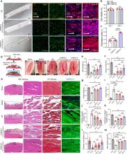
          Three-dimensional (3D) bioprinting is a highly effective technique for fabricating cell-loaded constructs in tissue engineering. However, the versatility of fabricating precise and complex cell-loaded hydrogels is limited owing to the poor crosslinking ability of cell-containing hydrogels. Herein, we propose an optic-fiber-assisted bioprinting (OAB) process to efficiently crosslink methacrylated hydrogels. By selecting appropriate processing conditions for the photo-crosslinking technique, we fabricated biofunctional cell-laden structures including methacrylated gelatin (Gelma), collagen, and decellularized extracellular matrix. To apply the method to skeletal muscle regeneration, cell-laden Gelma constructs were processed with a functional nozzle having a topographical cue and an OAB process that could induce a uniaxial alignment of C2C12 and human adipose stem cells (hASCs). Significantly higher degrees of cell alignment and myogenic activities in the cell-laden Gelma structure were observed compared with those in the cell construct that was printed using a conventional crosslinking method. Moreover, an in vivo regenerative potential was observed in volumetric muscle defects in a mouse model. The hASC-laden construct significantly induced greater muscle regeneration than the cell construct without topographical cues. Based on the results, the newly designed bioprinting process can prove to be highly effective in fabricating biofunctional cell-laden constructs for various tissue engineering applications.

          Related collections

          Most cited references49

          • Record: found
          • Abstract: found
          • Article: not found

          Biomimetic 4D printing.

          Shape-morphing systems can be found in many areas, including smart textiles, autonomous robotics, biomedical devices, drug delivery and tissue engineering. The natural analogues of such systems are exemplified by nastic plant motions, where a variety of organs such as tendrils, bracts, leaves and flowers respond to environmental stimuli (such as humidity, light or touch) by varying internal turgor, which leads to dynamic conformations governed by the tissue composition and microstructural anisotropy of cell walls. Inspired by these botanical systems, we printed composite hydrogel architectures that are encoded with localized, anisotropic swelling behaviour controlled by the alignment of cellulose fibrils along prescribed four-dimensional printing pathways. When combined with a minimal theoretical framework that allows us to solve the inverse problem of designing the alignment patterns for prescribed target shapes, we can programmably fabricate plant-inspired architectures that change shape on immersion in water, yielding complex three-dimensional morphologies.
            Bookmark
            • Record: found
            • Abstract: found
            • Article: not found

            Synthesis, properties, and biomedical applications of gelatin methacryloyl (GelMA) hydrogels.

            Gelatin methacryloyl (GelMA) hydrogels have been widely used for various biomedical applications due to their suitable biological properties and tunable physical characteristics. GelMA hydrogels closely resemble some essential properties of native extracellular matrix (ECM) due to the presence of cell-attaching and matrix metalloproteinase responsive peptide motifs, which allow cells to proliferate and spread in GelMA-based scaffolds. GelMA is also versatile from a processing perspective. It crosslinks when exposed to light irradiation to form hydrogels with tunable mechanical properties. It can also be microfabricated using different methodologies including micromolding, photomasking, bioprinting, self-assembly, and microfluidic techniques to generate constructs with controlled architectures. Hybrid hydrogel systems can also be formed by mixing GelMA with nanoparticles such as carbon nanotubes and graphene oxide, and other polymers to form networks with desired combined properties and characteristics for specific biological applications. Recent research has demonstrated the proficiency of GelMA-based hydrogels in a wide range of tissue engineering applications including engineering of bone, cartilage, cardiac, and vascular tissues, among others. Other applications of GelMA hydrogels, besides tissue engineering, include fundamental cell research, cell signaling, drug and gene delivery, and bio-sensing.
              Bookmark
              • Record: found
              • Abstract: found
              • Article: not found

              A 3D bioprinting system to produce human-scale tissue constructs with structural integrity

              A challenge for tissue engineering is producing three-dimensional (3D), vascularized cellular constructs of clinically relevant size, shape and structural integrity. We present an integrated tissue-organ printer (ITOP) that can fabricate stable, human-scale tissue constructs of any shape. Mechanical stability is achieved by printing cell-laden hydrogels together with biodegradable polymers in integrated patterns and anchored on sacrificial hydrogels. The correct shape of the tissue construct is achieved by representing clinical imaging data as a computer model of the anatomical defect and translating the model into a program that controls the motions of the printer nozzles, which dispense cells to discrete locations. The incorporation of microchannels into the tissue constructs facilitates diffusion of nutrients to printed cells, thereby overcoming the diffusion limit of 100-200 μm for cell survival in engineered tissues. We demonstrate capabilities of the ITOP by fabricating mandible and calvarial bone, cartilage and skeletal muscle. Future development of the ITOP is being directed to the production of tissues for human applications and to the building of more complex tissues and solid organs.
                Bookmark

                Author and article information

                Contributors
                dryu@gist.ac.kr
                gkimbme@skku.edu
                Journal
                NPJ Regen Med
                NPJ Regen Med
                NPJ Regenerative Medicine
                Nature Publishing Group UK (London )
                2057-3995
                31 March 2023
                31 March 2023
                2023
                : 8
                : 18
                Affiliations
                [1 ]GRID grid.264381.a, ISNI 0000 0001 2181 989X, Department of Precision Medicine, , Sungkyunkwan University School of Medicine, ; Suwon, Republic of Korea
                [2 ]GRID grid.222754.4, ISNI 0000 0001 0840 2678, Department of Biotechnology and Bioinformatics, , Korea University, ; Sejong, Republic of Korea
                [3 ]GRID grid.264381.a, ISNI 0000 0001 2181 989X, Department of Molecular Cell Biology, , Sungkyunkwan University School of Medicine, ; Suwon, Republic of Korea
                [4 ]GRID grid.61221.36, ISNI 0000 0001 1033 9831, Department of Biomedical Science and Engineering, , Gwangju Institute of Science and Technology, ; Gwangju, Republic of Korea
                [5 ]GRID grid.264381.a, ISNI 0000 0001 2181 989X, Department of Biophysics, Institute of Quantum Biophysics, , Sungkyunkwan University, ; Suwon, Republic of Korea
                Author information
                http://orcid.org/0000-0001-5905-6760
                http://orcid.org/0000-0002-2965-2171
                Article
                292
                10.1038/s41536-023-00292-5
                10066283
                37002225
                5e947907-4710-4e4e-88b4-ca6f335a28b6
                © The Author(s) 2023

                Open Access This article is licensed under a Creative Commons Attribution 4.0 International License, which permits use, sharing, adaptation, distribution and reproduction in any medium or format, as long as you give appropriate credit to the original author(s) and the source, provide a link to the Creative Commons license, and indicate if changes were made. The images or other third party material in this article are included in the article’s Creative Commons license, unless indicated otherwise in a credit line to the material. If material is not included in the article’s Creative Commons license and your intended use is not permitted by statutory regulation or exceeds the permitted use, you will need to obtain permission directly from the copyright holder. To view a copy of this license, visit http://creativecommons.org/licenses/by/4.0/.

                History
                : 2 July 2022
                : 17 March 2023
                Categories
                Article
                Custom metadata
                © The Author(s) 2023

                tissue engineering,translational research
                tissue engineering, translational research

                Comments

                Comment on this article