12
views
0
recommends
+1 Recommend
0 collections
    0
    shares
      • Record: found
      • Abstract: found
      • Article: found
      Is Open Access

      Circulating exosomal microRNAs reveal the mechanism of Fructus Meliae Toosendan-induced liver injury in mice

      research-article
      , , , ,
      Scientific Reports
      Nature Publishing Group UK

      Read this article at

      Bookmark
          There is no author summary for this article yet. Authors can add summaries to their articles on ScienceOpen to make them more accessible to a non-specialist audience.

          Abstract

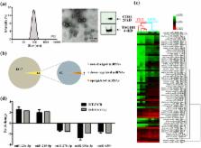
          The toxicological mechanisms of liver injury caused by most traditional Chinese medicine (TCM) remain largely unknown. Due to the unique features, exosomal microRNAs (miRNAs) are currently attracting major interests to provide further insights into toxicological mechanisms. Thus, taking Fructus Meliae Toosendan as an example of hepatoxic TCM, this study aimed to elucidate its hepatotoxicity mechanisms through profiling miRNAs in circulating exosomes of Fructus Meliae Toosendan water extract (FMT)-exposed mice. Biological pathway analysis of the 64 differentially expressed exosomal miRNAs (DEMs) showed that hepatic dysfunction induced by FMT likely related to apoptosis, mitochondrial dysfunction, and cell cycle dysregulation. Integrated analysis of serum exosomal DEMs and hepatic differentially expressed mRNAs further enriched oxidative stress and apoptosis related pathways. In vitro validation studies for omics results suggested that FMT-induced DNA damage was mediated by generating intracellular reactive oxygen species, leading to cell apoptosis through p53-dependent mitochondrial damage and S-phase arrest. Nrf2-mediated antioxidant response was activated to protect liver cells. Moreover, serum exosomal miR-370-3p, the most down-regulated miRNA involving in these pathways, might be the momentous event in aggravating cytotoxic effect of FMT by elevating p21 and Cyclin E. In conclusion, circulating exosomal miRNAs profiling could contribute to deepen the understanding of TCM-induced hepatotoxicity.

          Related collections

          Most cited references39

          • Record: found
          • Abstract: found
          • Article: not found
          Is Open Access

          The Nrf2-antioxidant response element signaling pathway and its activation by oxidative stress.

          A major mechanism in the cellular defense against oxidative or electrophilic stress is activation of the Nrf2-antioxidant response element signaling pathway, which controls the expression of genes whose protein products are involved in the detoxication and elimination of reactive oxidants and electrophilic agents through conjugative reactions and by enhancing cellular antioxidant capacity. At the molecular level, however, the regulatory mechanisms involved in mediating Nrf2 activation are not fully understood. It is well established that Nrf2 activity is controlled, in part, by the cytosolic protein Keap1, but the nature of this pathway and the mechanisms by which Keap1 acts to repress Nrf2 activity remain to be fully characterized and are the topics of discussion in this minireview. In addition, a possible role of the Nrf2-antioxidant response element transcriptional pathway in neuroprotection will also be discussed.
            Bookmark
            • Record: found
            • Abstract: found
            • Article: not found

            Causes, clinical features, and outcomes from a prospective study of drug-induced liver injury in the United States.

            Idiosyncratic drug-induced liver injury (DILI) is among the most common causes of acute liver failure in the United States, accounting for approximately 13% of cases. A prospective study was begun in 2003 to recruit patients with suspected DILI and create a repository of biological samples for analysis. This report summarizes the causes, clinical features, and outcomes from the first 300 patients enrolled. Patients with suspected DILI were enrolled based on predefined criteria and followed up for at least 6 months. Patients with acetaminophen liver injury were excluded. DILI was caused by a single prescription medication in 73% of the cases, by dietary supplements in 9%, and by multiple agents in 18%. More than 100 different agents were associated with DILI; antimicrobials (45.5%) and central nervous system agents (15%) were the most common. Causality was considered to be definite in 32%, highly likely in 41%, probable in 14%, possible in 10%, and unlikely in 3%. Acute hepatitis C virus (HCV) infection was the final diagnosis in 4 of 9 unlikely cases. Six months after enrollment, 14% of patients had persistent laboratory abnormalities and 8% had died; the cause of death was liver related in 44%. DILI is caused by a wide array of medications, herbal supplements, and dietary supplements. Antibiotics are the single largest class of agents that cause DILI. Acute HCV infection should be excluded in patients with suspected DILI by HCV RNA testing. The overall 6-month mortality was 8%, but the majority of deaths were not liver related.
              Bookmark
              • Record: found
              • Abstract: found
              • Article: not found

              Mechanisms of hepatotoxicity.

              H Jaeschke (2002)
              This review addresses recent advances in specific mechanisms of hepatotoxicity. Because of its unique metabolism and relationship to the gastrointestinal tract, the liver is an important target of the toxicity of drugs, xenobiotics, and oxidative stress. In cholestatic disease, endogenously generated bile acids produce hepatocellular apoptosis by stimulating Fas translocation from the cytoplasm to the plasma membrane where self-aggregation occurs to trigger apoptosis. Kupffer cell activation and neutrophil infiltration extend toxic injury. Kupffer cells release reactive oxygen species (ROS), cytokines, and chemokines, which induce neutrophil extravasation and activation. The liver expresses many cytochrome P450 isoforms, including ethanol-induced CYP2E1. CYP2E1 generates ROS, activates many toxicologically important substrates, and may be the central pathway by which ethanol causes oxidative stress. In acetaminophen toxicity, nitric oxide (NO) scavenges superoxide to produce peroxynitrite, which then causes protein nitration and tissue injury. In inducible nitric oxide synthase (iNOS) knockout mice, nitration is prevented, but unscavenged superoxide production then causes toxic lipid peroxidation to occur instead. Microvesicular steatosis, nonalcoholic steatohepatitis (NASH), and cytolytic hepatitis involve mitochondrial dysfunction, including impairment of mitochondrial fatty acid beta-oxidation, inhibition of mitochondrial respiration, and damage to mitochondrial DNA. Induction of the mitochondrial permeability transition (MPT) is another mechanism causing mitochondrial failure, which can lead to necrosis from ATP depletion or caspase-dependent apoptosis if ATP depletion does not occur fully. Because of such diverse mechanisms, hepatotoxicity remains a major reason for drug withdrawal from pharmaceutical development and clinical use.
                Bookmark

                Author and article information

                Contributors
                luxy@zju.edu.cn
                fanxh@zju.edu.cn
                Journal
                Sci Rep
                Sci Rep
                Scientific Reports
                Nature Publishing Group UK (London )
                2045-2322
                12 February 2018
                12 February 2018
                2018
                : 8
                : 2832
                Affiliations
                ISNI 0000 0004 1759 700X, GRID grid.13402.34, Pharmaceutical Informatics Institute, College of Pharmaceutical Sciences, , Zhejiang University, ; Hangzhou, 310058 China
                Author information
                http://orcid.org/0000-0002-6336-3007
                Article
                21113
                10.1038/s41598-018-21113-6
                5809479
                29434260
                5bff40f8-a759-4516-8492-4bd13af7fa30
                © The Author(s) 2018

                Open Access This article is licensed under a Creative Commons Attribution 4.0 International License, which permits use, sharing, adaptation, distribution and reproduction in any medium or format, as long as you give appropriate credit to the original author(s) and the source, provide a link to the Creative Commons license, and indicate if changes were made. The images or other third party material in this article are included in the article’s Creative Commons license, unless indicated otherwise in a credit line to the material. If material is not included in the article’s Creative Commons license and your intended use is not permitted by statutory regulation or exceeds the permitted use, you will need to obtain permission directly from the copyright holder. To view a copy of this license, visit http://creativecommons.org/licenses/by/4.0/.

                History
                : 2 November 2017
                : 29 January 2018
                Categories
                Article
                Custom metadata
                © The Author(s) 2018

                Uncategorized
                Uncategorized

                Comments

                Comment on this article