48
views
0
recommends
+1 Recommend
0 collections
    0
    shares
      • Record: found
      • Abstract: found
      • Article: found
      Is Open Access

      Chondrocytes Transdifferentiate into Osteoblasts in Endochondral Bone during Development, Postnatal Growth and Fracture Healing in Mice

      research-article

      Read this article at

      Bookmark
          There is no author summary for this article yet. Authors can add summaries to their articles on ScienceOpen to make them more accessible to a non-specialist audience.

          Abstract

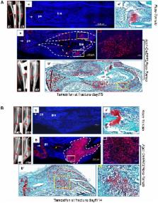
          One of the crucial steps in endochondral bone formation is the replacement of a cartilage matrix produced by chondrocytes with bone trabeculae made by osteoblasts. However, the precise sources of osteoblasts responsible for trabecular bone formation have not been fully defined. To investigate whether cells derived from hypertrophic chondrocytes contribute to the osteoblast pool in trabecular bones, we genetically labeled either hypertrophic chondrocytes by Col10a1-Cre or chondrocytes by tamoxifen-induced Agc1-CreERT2 using EGFP, LacZ or Tomato expression. Both Cre drivers were specifically active in chondrocytic cells and not in perichondrium, in periosteum or in any of the osteoblast lineage cells. These in vivo experiments allowed us to follow the fate of cells labeled in Col10a1-Cre or Agc1-CreERT2 -expressing chondrocytes. After the labeling of chondrocytes, both during prenatal development and after birth, abundant labeled non-chondrocytic cells were present in the primary spongiosa. These cells were distributed throughout trabeculae surfaces and later were present in the endosteum, and embedded within the bone matrix. Co-expression studies using osteoblast markers indicated that a proportion of the non-chondrocytic cells derived from chondrocytes labeled by Col10a1-Cre or by Agc1-CreERT2 were functional osteoblasts. Hence, our results show that both chondrocytes prior to initial ossification and growth plate chondrocytes before or after birth have the capacity to undergo transdifferentiation to become osteoblasts. The osteoblasts derived from Col10a1-expressing hypertrophic chondrocytes represent about sixty percent of all mature osteoblasts in endochondral bones of one month old mice. A similar process of chondrocyte to osteoblast transdifferentiation was involved during bone fracture healing in adult mice. Thus, in addition to cells in the periosteum chondrocytes represent a major source of osteoblasts contributing to endochondral bone formation in vivo.

          Author Summary

          During endochondral bone formation, which is responsible for the generation of most bones in mammals and many other species, osteoblasts deposit a bone-specific matrix on the surface of cartilage scaffolds made by chondrocytes and hypertrophic chondrocytes. It has long been thought that the terminally differentiated chondrocytes in this cartilage scaffold undergo cell death. Here we demonstrate that chondrocytes can transdifferentiate into osteoblasts and that these transdifferentiated osteoblasts represent a substantial fraction of the bone forming cells in mice, We also provide evidence that chondrocytes can transdifferentiate into osteoblasts during bone fracture repair, a process similar to endochondral bone formation.

          Related collections

          Most cited references24

          • Record: found
          • Abstract: found
          • Article: not found

          Wnt/beta-catenin signaling in mesenchymal progenitors controls osteoblast and chondrocyte differentiation during vertebrate skeletogenesis.

          Chondrocytes and osteoblasts are two primary cell types in the skeletal system that are differentiated from common mesenchymal progenitors. It is believed that osteoblast differentiation is controlled by distinct mechanisms in intramembranous and endochondral ossification. We have found that ectopic canonical Wnt signaling leads to enhanced ossification and suppression of chondrocyte formation. Conversely, genetic inactivation of beta-catenin, an essential component transducing the canonical Wnt signaling, causes ectopic formation of chondrocytes at the expense of osteoblast differentiation during both intramembranous and endochondral ossification. Moreover, inactivation of beta-catenin in mesenchymal progenitor cells in vitro causes chondrocyte differentiation under conditions allowing only osteoblasts to form. Our results demonstrate that beta-catenin is essential in determining whether mesenchymal progenitors will become osteoblasts or chondrocytes regardless of regional locations or ossification mechanisms. Controlling Wnt/beta-catenin signaling is a common molecular mechanism underlying chondrocyte and osteoblast differentiation and specification of intramembranous and endochondral ossification.
            Bookmark
            • Record: found
            • Abstract: found
            • Article: not found

            Osteoblast precursors, but not mature osteoblasts, move into developing and fractured bones along with invading blood vessels.

            During endochondral bone development, the first osteoblasts differentiate in the perichondrium surrounding avascular cartilaginous rudiments; the source of trabecular osteoblasts inside the later bone is, however, unknown. Here, we generated tamoxifen-inducible transgenic mice bred to Rosa26R-LacZ reporter mice to follow the fates of stage-selective subsets of osteoblast lineage cells. Pulse-chase studies showed that osterix-expressing osteoblast precursors, labeled in the perichondrium prior to vascular invasion of the cartilage, give rise to trabecular osteoblasts, osteocytes, and stromal cells inside the developing bone. Throughout the translocation, some precursors were found to intimately associate with invading blood vessels, in pericyte-like fashion. A similar coinvasion occurs during endochondral healing of bone fractures. In contrast, perichondrial mature osteoblasts did not exhibit perivascular localization and remained in the outer cortex of developing bones. These findings reveal the specific involvement of immature osteoblast precursors in the coupled vascular and osteogenic transformation essential to endochondral bone development and repair. 2010 Elsevier Inc. All rights reserved.
              Bookmark
              • Record: found
              • Abstract: found
              • Article: not found

              Fracture healing as a post-natal developmental process: molecular, spatial, and temporal aspects of its regulation.

              Fracture healing is a specialized post-natal repair process that recapitulates aspects of embryological skeletal development. While many of the molecular mechanisms that control cellular differentiation and growth during embryogenesis recur during fracture healing, these processes take place in a post-natal environment that is unique and distinct from those which exist during embryogenesis. This Prospect Article will highlight a number of central biological processes that are believed to be crucial in the embryonic differentiation and growth of skeletal tissues and review the functional role of these processes during fracture healing. Specific aspects of fracture healing that will be considered in relation to embryological development are: (1) the anatomic structure of the fracture callus as it evolves during healing; (2) the origins of stem cells and morphogenetic signals that facilitate the repair process; (3) the role of the biomechanical environment in controlling cellular differentiation during repair; (4) the role of three key groups of soluble factors, pro-inflammatory cytokines, the TGF-beta superfamily, and angiogenic factors, during repair; and (5) the relationship of the genetic components that control bone mass and remodeling to the mechanisms that control skeletal tissue repair in response to fracture. Copyright 2003 Wiley-Liss, Inc.
                Bookmark

                Author and article information

                Contributors
                Role: Editor
                Journal
                PLoS Genet
                PLoS Genet
                plos
                plosgen
                PLoS Genetics
                Public Library of Science (San Francisco, USA )
                1553-7390
                1553-7404
                December 2014
                4 December 2014
                : 10
                : 12
                : e1004820
                Affiliations
                [1 ]Department of Genetics, The University of Texas MD Anderson Cancer Center, Houston, Texas, United States of America
                [2 ]Department of Experimental Medicine 1, Nikolaus-Fiebiger-Center of Molecular Medicine, University of Erlangen-Nuremberg, Erlangen, Germany
                [3 ]Department of Veterinary Medicine & Surgery, The University of Texas MD Anderson Cancer Center, Houston, Texas, United States of America
                Children's Hospital Boston and Harvard Medical School, Howard Hughes Medical Institute, United States of America
                Author notes

                The authors have declared that no competing interests exist.

                Conceived and designed the experiments: XZ BdC. Performed the experiments: XZ WN. Analyzed the data: XZ BdC. Contributed reagents/materials/analysis tools: KvdM SH HA. Wrote the paper: XZ BdC.

                Article
                PGENETICS-D-12-02047
                10.1371/journal.pgen.1004820
                4256265
                25474590
                5bbc3249-8335-4c16-bcef-c9626dedd533
                Copyright @ 2014

                This is an open-access article distributed under the terms of the Creative Commons Attribution License, which permits unrestricted use, distribution, and reproduction in any medium, provided the original author and source are credited.

                History
                : 10 August 2012
                : 14 October 2014
                Page count
                Pages: 20
                Funding
                The work is supported by NIH# 2RO1 AR049072-11A1 and 2RO1 AR049072-06 (BdC), the Rolanette and Berdon Lawrence Bone Research Award (XZ 2013-2014), and the Deutsche Forschungsgemeinschaft, grant nr. MA534/23-2 (KvdM). The funders had no role in study design, data collection and analysis, decision to publish, or preparation of the manuscript.
                Categories
                Research Article
                Biology and Life Sciences

                Genetics
                Genetics

                Comments

                Comment on this article