20
views
0
recommends
+1 Recommend
1 collections
    0
    shares
      • Record: found
      • Abstract: found
      • Article: found
      Is Open Access

      Dental anomalies in an orthodontic patient population with maxillary lateral incisor agenesis

      research-article

      Read this article at

      ScienceOpenPublisherPMC
      Bookmark
          There is no author summary for this article yet. Authors can add summaries to their articles on ScienceOpen to make them more accessible to a non-specialist audience.

          ABSTRACT

          Introduction:

          The purpose of this study was to evaluate the prevalence of dental anomalies in a subpopulation of orthodontic patients with agenesis of maxillary lateral incisors (MLI).

          Methods:

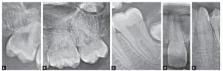
          The material of the present study included the records of the 1964 orthodontic patients. Panoramic radiographs and dental casts were used to analyze other associated eight dental anomalies, including agenesis of other teeth, dens invaginatus, dens evaginatus, peg shaped MLI, taurodontism, pulp stone, root dilaceration and maxillary canine impaction.

          Results:

          Out of the 1964 patients examined, 90 were found to have agenesis of MLI, representing a prevalence of 4.6%. The most commonly found associated-anomalies were agenesis of other teeth (23.3%), peg-shaped MLIs (15.6%), taurodontism (42.2%), and dilacerated teeth (18.9%).

          Conclusion:

          Permanent tooth agenesis, taurodontism, peg-shaped maxillary lateral incisor, and root dilacerations are frequently associated with maxillary lateral incisor agenesis.

          RESUMO

          Objetivo:

          o objetivo do presente estudo foi avaliar a prevalência de anomalias dentárias em uma subpopulação de pacientes ortodônticos com agenesia de incisivos laterais superiores (ILS).

          Material e Métodos:

          o material do presente estudo incluiu os registros de 1964 pacientes ortodônticos. Radiografias panorâmicas e modelos de estudo foram usados para analisar outras anomalias dentárias associadas, incluindo a agenesia de outros dentes, dens invaginatus, dens evaginatus, ILS conoides, taurodontismo, calcificação pulpar, dilaceração radicular e impacção do canino superior.

          Resultados:

          dos 1964 pacientes examinados, constatou-se que 90 tinham agenesia do ILS, o que representa uma prevalência de 4,6%. As anomalias associadas mais comumente encontradas foram a agenesia de outros dentes (23,3%), ILS conoides (15,6%), taurodontismo (42,2%) e dentes com dilaceração (18,9%).

          Conclusão:

          a agenesia de dentes permanentes, o taurodontismo, incisivos laterais superiores conoides e dilaceração radicular estão frequentemente associados à agenesia de incisivos laterais superiores.

          Related collections

          Most cited references77

          • Record: found
          • Abstract: found
          • Article: not found

          A meta-analysis of the prevalence of dental agenesis of permanent teeth.

          To gain more insight into the prevalence of dental agenesis. Data from Caucasian populations in North America, Australia and Europe were included in a meta-analysis. For the prevalence of African American, Chinese and Arab groups only indications could be reported because of a limited number of studies. Agenesis differs by continent and gender: the prevalence for both sexes was higher in Europe (males 4.6%; females 6.3%) and Australia (males 5.5%; females 7.6%) than for North American Caucasians (males 3.2%; females 4.6%). In addition, the prevalence of dental agenesis in females was 1.37 times higher than in males. The mandibular second premolar was the most affected tooth, followed by the maxillary lateral incisor and the maxillary second premolar. The occurrence of dental agenesis was divided into three main groups: common (P2(i) > I2(s) > P2(s)), less common (I1(i) > I2(i) & P1(s) > C(s) & M2(i)) and rare (M2(s) & M1(s) > C(i) > M1(i) & I1(s)). Unilateral occurrence of dental agenesis is more common than bilateral occurrence. However, bilateral agenesis of maxillary lateral incisors is more common than unilateral agenesis. The overall prevalence of agenesis in the maxilla is comparable with that in the mandible, but a marked difference was found between both jaws regarding tooth type. Absence of one or two permanent teeth is found in 83% of the subjects with dental agenesis. A practical application of the results of the meta-analysis is the estimation of dental treatment need. Copyright Blackwell Munksgaard, 2004
            Bookmark
            • Record: found
            • Abstract: found
            • Article: not found

            Prevalence and distribution of dental anomalies in orthodontic patients.

            The purpose of this study was to determine the prevalence of developmental dental anomalies in the Turkish population. The study was based on the dental casts, intraoral photographs, and panoramic radiographs of 3043 Turkish children (1658 girls, 1385 boys) who had orthodontic treatment at the Department of Orthodontics at the University of Ankara between 1978 and 2003. These patients were examined for 8 developmental dental anomalies: fusion, gemination, microdontia, macrodontia, oligodontia, hypodontia, hyperdontia, and amelogenesis imperfecta. The percentages of these anomalies were assessed in the whole group, and the sexes were compared. It was found that 5.46% of the total group had at least 1 developmental dental anomaly. The distribution by sex was 70 boys (5.05%) and 96 girls (5.79%). Hypodontia is the most common developmental dental anomaly in the Turkish population, followed by microdontia.
              Bookmark
              • Record: found
              • Abstract: found
              • Article: not found

              Genetic basis of tooth agenesis.

              Tooth agenesis or hypodontia, failure to develop all normally developing teeth, is one of the most common developmental anomalies in man. Common forms, including third molar agenesis and hypodontia of one or more of the incisors and premolars, constitute the great majority of cases. They typically affect those teeth that develop latest in each tooth class and these teeth are also most commonly affected in more severe and rare types of tooth agenesis. Specific vulnerability of the last developing teeth suggests that agenesis reflects quantitative defects during dental development. So far molecular genetics has revealed the genetic background of only rare forms of tooth agenesis. Mutations in MSX1, PAX9, AXIN2 and EDA have been identified in familial severe agenesis (oligodontia) and mutations in many other genes have been identified in syndromes in which tooth agenesis is a regular feature. Heterozygous loss of function mutations in many genes reduce the gene dose, whereas e.g. in hypohidrotic ectodermal dysplasia (EDA) the complete inactivation of the partially redundant signaling pathway reduces the signaling centers. Although these mechanisms involve quantitative disturbances, the phenotypes associated with mutations in different genes indicate that in addition to an overall reduction of odontogenic potential, tooth class-specific and more complex mechanisms are also involved. Although several of the genes so far identified in rare forms of tooth agenesis are being studied as candidate genes of common third molar agenesis and incisor and premolar hypodontia, it is plausible that novel genes that contribute to these phenotypes will also become identified. (c) 2009 Wiley-Liss, Inc.
                Bookmark

                Author and article information

                Journal
                Dental Press J Orthod
                Dental Press J Orthod
                dpjo
                Dental Press Journal of Orthodontics
                Dental Press International
                2176-9451
                2177-6709
                Nov-Dec 2016
                Nov-Dec 2016
                : 21
                : 6
                : 98-102
                Affiliations
                [1 ]Research Assistant, Department of Endodontics, Faculty of Dentistry, Ordu University, Ordu, Turkey.
                [2 ]Assistant Professor, Department of Endodontics, Faculty of Dentistry, Ordu University, Ordu, Turkey.
                [3 ]Assistant Professor, Department of Orthodontics, Faculty of Dentistry, Ordu University, Ordu, Turkey.
                [4 ]Research Assistant, Department of Orthodontics, Faculty of Dentistry, Ordu University, Ordu, Turkey.
                Author notes
                Contact address: Suleyman Kutalmış Buyuk E-mail: skbuyuk@ 123456gmail.com

                Author contributions: Conception/design of the study: SKB, YAB, FC. Acquisition, analysis or interpretation: YAB, BB, MC, SKB. Wrote the article: SKB, EBC. Critical revision of the article: SKB, YAB. Final approval of the article: SKB, EBC.

                Article
                10.1590/2177-6709.21.6.098-102.oar
                5278939
                28125145
                5bb0a190-34ec-4195-97a2-0f19cd47cab0

                This is an open-access article distributed under the terms of the Creative Commons Attribution License

                History
                : 18 March 2016
                : 20 July 2016
                Page count
                Figures: 1, Tables: 2, Equations: 0, References: 44, Pages: 5
                Categories
                Original Article

                dental anomalies,hypodontia,panoramic radiograph
                dental anomalies, hypodontia, panoramic radiograph

                Comments

                Comment on this article