10
views
0
recommends
+1 Recommend
0 collections
    0
    shares
      • Record: found
      • Abstract: found
      • Article: not found

      Crystal structures of Ryanodine Receptor SPRY1 and tandem-repeat domains reveal a critical FKBP12 binding determinant

      research-article

      Read this article at

      ScienceOpenPublisherPMC
      Bookmark
          There is no author summary for this article yet. Authors can add summaries to their articles on ScienceOpen to make them more accessible to a non-specialist audience.

          Abstract

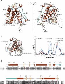
          Ryanodine receptors (RyRs) form calcium release channels located in the membranes of the sarcoplasmic and endoplasmic reticulum. RyRs play a major role in excitation-contraction coupling and other Ca 2+-dependent signaling events, and consist of several globular domains that together form a large assembly. Here we describe the crystal structures of the SPRY1 and tandem-repeat domains at 1.2 – 1.5Å resolution, which reveal several structural elements not detected in recent cryo-EM reconstructions of RyRs. The cryo-EM studies disagree on the position of SPRY domains, which had been proposed based on homology modeling. Computational docking of the crystal structures, combined with FRET studies, show that the SPRY1 domain is located next to FK506-Binding Protein (FKBP). Molecular dynamics flexible fitting and mutagenesis experiments suggest a hydrophobic cluster within SPRY1 that is crucial for FKBP binding. A RyR1 disease mutation, N760D, appears to directly impact FKBP binding through interfering with SPRY1 folding.

          Related collections

          Most cited references53

          • Record: found
          • Abstract: found
          • Article: not found

          Flexible fitting of atomic structures into electron microscopy maps using molecular dynamics.

          A novel method to flexibly fit atomic structures into electron microscopy (EM) maps using molecular dynamics simulations is presented. The simulations incorporate the EM data as an external potential added to the molecular dynamics force field, allowing all internal features present in the EM map to be used in the fitting process, while the model remains fully flexible and stereochemically correct. The molecular dynamics flexible fitting (MDFF) method is validated for available crystal structures of protein and RNA in different conformations; measures to assess and monitor the fitting process are introduced. The MDFF method is then used to obtain high-resolution structures of the E. coli ribosome in different functional states imaged by cryo-EM.
            Bookmark
            • Record: found
            • Abstract: found
            • Article: not found

            Ryanodine receptor oxidation causes intracellular calcium leak and muscle weakness in aging.

            Age-related loss of muscle mass and force (sarcopenia) contributes to disability and increased mortality. Ryanodine receptor 1 (RyR1) is the skeletal muscle sarcoplasmic reticulum calcium release channel required for muscle contraction. RyR1 from aged (24 months) rodents was oxidized, cysteine-nitrosylated, and depleted of the channel-stabilizing subunit calstabin1, compared to RyR1 from younger (3-6 months) adults. This RyR1 channel complex remodeling resulted in "leaky" channels with increased open probability, leading to intracellular calcium leak in skeletal muscle. Similarly, 6-month-old mice harboring leaky RyR1-S2844D mutant channels exhibited skeletal muscle defects comparable to 24-month-old wild-type mice. Treating aged mice with S107 stabilized binding of calstabin1 to RyR1, reduced intracellular calcium leak, decreased reactive oxygen species (ROS), and enhanced tetanic Ca(2+) release, muscle-specific force, and exercise capacity. Taken together, these data indicate that leaky RyR1 contributes to age-related loss of muscle function. Copyright © 2011 Elsevier Inc. All rights reserved.
              Bookmark
              • Record: found
              • Abstract: found
              • Article: not found

              Structure of the rabbit ryanodine receptor RyR1 at near-atomic resolution

              The ryanodine receptors (RyRs) are high-conductance intracellular Ca2+ channels that play a pivotal role in the excitation-contraction coupling of skeletal and cardiac muscles. RyRs are the largest known ion channels, with a homotetrameric organization and approximately 5000 residues in each protomer. Here we report the structure of the rabbit RyR1 in complex with its modulator FKBP12 at an overall resolution of 3.8 Å, determined by single-particle electron cryo-microscopy. Three previously uncharacterized domains, named Central, Handle, and Helical domains, display the armadillo repeat fold. These domains, together with the amino-terminal domain, constitute a network of superhelical scaffold for binding and propagation of conformational changes. The channel domain exhibits the voltage-gated ion channel superfamily fold with distinct features. A negative charge-enriched hairpin loop connecting S5 and the pore helix is positioned above the entrance to the selectivity filter vestibule. The four elongated S6 segments form a right-handed helical bundle that closes the pore at the cytoplasmic border of the membrane. Allosteric regulation of the pore by the cytoplasmic domains is mediated through extensive interactions between the Central domains and the channel domain. These structural features explain high ion conductance by RyRs and the long-range allosteric regulation of channel activities.
                Bookmark

                Author and article information

                Journal
                101528555
                37539
                Nat Commun
                Nat Commun
                Nature communications
                2041-1723
                10 July 2015
                06 August 2015
                2015
                06 February 2016
                : 6
                : 7947
                Affiliations
                [1 ] Department of Biochemistry and Molecular Biology, The Life Sciences Centre, University of British Columbia, 2350 Health Sciences Mall, V6T 1Z3, Vancouver, Canada
                [2 ] Department of Biochemistry, Molecular Biology and Biophysics, University of Minnesota, 312 Church St SE, Minneapolis, MN 55455, USA
                [3 ]Department of Anesthesia, Perioperative and Pain Medicine, Brigham and Women's Hospital,75 Francis Street, Boston, MA 02115, USA.
                Author notes
                Article
                NIHMS704401
                10.1038/ncomms8947
                4530471
                26245150
                5afa2bec-d4b8-4fdc-9940-18345c6326e8

                Users may view, print, copy, and download text and data-mine the content in such documents, for the purposes of academic research, subject always to the full Conditions of use: http://www.nature.com/authors/editorial_policies/license.html#terms

                History
                Categories
                Article

                Uncategorized
                Uncategorized

                Comments

                Comment on this article