17
views
0
recommends
+1 Recommend
0 collections
    0
    shares
      • Record: found
      • Abstract: found
      • Article: found
      Is Open Access

      Prediction of Pseudoprogression versus Progression using Machine Learning Algorithm in Glioblastoma

      research-article

      Read this article at

      Bookmark
          There is no author summary for this article yet. Authors can add summaries to their articles on ScienceOpen to make them more accessible to a non-specialist audience.

          Abstract

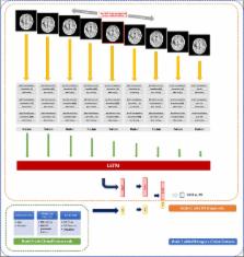
          We aimed to investigate the feasibility of machine learning (ML) algorithm to distinguish pseudoprogression (PsPD) from progression (PD) in patients with glioblastoma (GBM). We recruited the patients diagnosed as primary GBM who received gross total resection (GTR) and concurrent chemoradiotherapy in two institutions from April 2010 to April 2017 and presented suspicious contrast-enhanced lesion on brain magnetic resonance imaging (MRI) during follow-up. Patients from two institutions were allocated to training (N = 59) and testing (N = 19) datasets, respectively. We developed a convolutional neural network combined with a long short-term memory ML structure. MRI data, which was 9 axial post-contrast T1-weighted images in our study, and clinical features were incorporated (Model 1). In the testing set, the trained Model 1 resulted in AUC of 0.83, AUPRC of 0.87, and F1-score of 0.74 using optimal threshold. The performance was superior to that of Model 2 (CNN-LSTM model with MRI data alone) and Model 3 (random forest model with clinical feature alone). The developed algorithm involving MRI data and clinical features could help making decision during follow-up of patients with GBM treated with GTR and concurrent CCRT.

          Related collections

          Most cited references22

          • Record: found
          • Abstract: not found
          • Book Chapter: not found

          Mitosis Detection in Breast Cancer Histology Images with Deep Neural Networks

            Bookmark
            • Record: found
            • Abstract: found
            • Article: not found

            MGMT promoter methylation status can predict the incidence and outcome of pseudoprogression after concomitant radiochemotherapy in newly diagnosed glioblastoma patients.

            Standard therapy for glioblastoma (GBM) is temozolomide (TMZ) administration, initially concurrent with radiotherapy (RT), and subsequently as maintenance therapy. The radiologic images obtained in this setting can be difficult to interpret since they may show radiation-induced pseudoprogression (psPD) rather than disease progression. Patients with histologically confirmed GBM underwent radiotherapy plus continuous daily temozolomide (75 mg/m(2)/d), followed by 12 maintenance temozolomide cycles (150 to 200 mg/m(2) for 5 days every 28 days) if magnetic resonance imaging (MRI) showed no enhancement suggesting a tumor; otherwise, chemotherapy was delivered until complete response or unequivocal progression. The first MRI scan was performed 1 month after completing combined chemoradiotherapy. In 103 patients (mean age, 52 years [range 20 to 73 years]), total resection, subtotal resection, and biopsy were obtained in 51, 51, and 1 cases, respectively. MGMT promoter was methylated in 36 patients (35%) and unmethylated in 67 patients (65%). Lesion enlargement, evidenced at the first MRI scan in 50 of 103 patients, was subsequently classified as psPD in 32 patients and early disease progression in 18 patients. PsPD was recorded in 21 (91%) of 23 methylated MGMT promoter and 11 (41%) of 27 unmethylated MGMT promoter (P = .0002) patients. MGMT status (P = .001) and psPD detection (P = .045) significantly influenced survival. PsPD has a clinical impact on chemotherapy-treated GBM, as it may express the glioma killing effects of treatment and is significantly correlated with MGMT status. Improvement in the early recognition of psPD patterns and knowledge of mechanisms underlying this phenomenon are crucial to eliminating biases in evaluating the results of clinical trials and guaranteeing effective treatment.
              Bookmark
              • Record: found
              • Abstract: found
              • Article: not found

              Long-Term Recurrent Convolutional Networks for Visual Recognition and Description.

              Models based on deep convolutional networks have dominated recent image interpretation tasks; we investigate whether models which are also recurrent are effective for tasks involving sequences, visual and otherwise. We describe a class of recurrent convolutional architectures which is end-to-end trainable and suitable for large-scale visual understanding tasks, and demonstrate the value of these models for activity recognition, image captioning, and video description. In contrast to previous models which assume a fixed visual representation or perform simple temporal averaging for sequential processing, recurrent convolutional models are "doubly deep" in that they learn compositional representations in space and time. Learning long-term dependencies is possible when nonlinearities are incorporated into the network state updates. Differentiable recurrent models are appealing in that they can directly map variable-length inputs (e.g., videos) to variable-length outputs (e.g., natural language text) and can model complex temporal dynamics; yet they can be optimized with backpropagation. Our recurrent sequence models are directly connected to modern visual convolutional network models and can be jointly trained to learn temporal dynamics and convolutional perceptual representations. Our results show that such models have distinct advantages over state-of-the-art models for recognition or generation which are separately defined or optimized.
                Bookmark

                Author and article information

                Contributors
                inah228@snu.ac.kr
                Journal
                Sci Rep
                Sci Rep
                Scientific Reports
                Nature Publishing Group UK (London )
                2045-2322
                21 August 2018
                21 August 2018
                2018
                : 8
                : 12516
                Affiliations
                [1 ]ISNI 0000 0001 0302 820X, GRID grid.412484.f, Department of Radiation Oncology, , Seoul National University Hospital, ; Seoul, Korea
                [2 ]ISNI 0000 0004 0647 3378, GRID grid.412480.b, Department of Radiation Oncology, , Seoul National University Bundang Hospital, ; Seongnamsi, Korea
                [3 ]ISNI 0000 0004 0470 5905, GRID grid.31501.36, Institute of Radiation Medicine, Cancer Research Institute, , Seoul National University College of Medicine, ; Seoul, Korea
                Article
                31007
                10.1038/s41598-018-31007-2
                6104063
                30131513
                5a8312be-c56c-4153-bdd9-e00b85d2f44f
                © The Author(s) 2018

                Open Access This article is licensed under a Creative Commons Attribution 4.0 International License, which permits use, sharing, adaptation, distribution and reproduction in any medium or format, as long as you give appropriate credit to the original author(s) and the source, provide a link to the Creative Commons license, and indicate if changes were made. The images or other third party material in this article are included in the article’s Creative Commons license, unless indicated otherwise in a credit line to the material. If material is not included in the article’s Creative Commons license and your intended use is not permitted by statutory regulation or exceeds the permitted use, you will need to obtain permission directly from the copyright holder. To view a copy of this license, visit http://creativecommons.org/licenses/by/4.0/.

                History
                : 16 March 2018
                : 9 August 2018
                Funding
                Funded by: This work was supported by Seoul National University Big Data Institute via The Data Science Research Project 2017 and SNUBH Research Fund (#14-2018-003) to In Ah Kim.
                Categories
                Article
                Custom metadata
                © The Author(s) 2018

                Uncategorized
                Uncategorized

                Comments

                Comment on this article