46
views
0
recommends
+1 Recommend
0 collections
    0
    shares
      • Record: found
      • Abstract: found
      • Article: found
      Is Open Access

      C-C Chemokine Receptor 2 (CCR2) Regulates the Hepatic Recruitment of Myeloid Cells That Promote Obesity-Induced Hepatic Steatosis

      research-article

      Read this article at

      Bookmark
          There is no author summary for this article yet. Authors can add summaries to their articles on ScienceOpen to make them more accessible to a non-specialist audience.

          Abstract

          OBJECTIVE

          Obesity induces a program of systemic inflammation that is implicated in the development of many of its clinical sequelae. Hepatic inflammation is a feature of obesity-induced liver disease, and our previous studies demonstrated reduced hepatic steatosis in obese mice deficient in the C-C chemokine receptor 2 (CCR2) that regulates myeloid cell recruitment. This suggests that a myeloid cell population is recruited to the liver in obesity and contributes to nonalcoholic fatty liver disease.

          RESEARCH DESIGN AND METHODS

          We used fluorescence-activated cell sorting to measure hepatic leukocyte populations in genetic and diet forms of murine obesity. We characterized in vivo models that increase and decrease an obesity-regulated CCR2-expressing population of hepatic leukocytes. Finally, using an in vitro co-culture system, we measured the ability of these cells to modulate a hepatocyte program of lipid metabolism.

          RESULTS

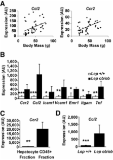
          We demonstrate that obesity activates hepatocyte expression of C-C chemokine ligand 2 (CCL2/MCP-1) leading to hepatic recruitment of CCR2 + myeloid cells that promote hepatosteatosis. The quantity of these cells correlates with body mass and in obese mice represents the second largest immune cell population in the liver. Hepatic expression of CCL2 increases their recruitment and in the presence of dietary fat induces hepatosteatosis. These cells activate hepatic transcription of genes responsible for fatty acid esterification and steatosis.

          CONCLUSIONS

          Obesity induces hepatic recruitment of a myeloid cell population that promotes hepatocyte lipid storage. These findings demonstrate that recruitment of myeloid cells to metabolic tissues is a common feature of obesity, not limited to adipose tissue.

          Related collections

          Most cited references24

          • Record: found
          • Abstract: found
          • Article: not found

          MCP-1 contributes to macrophage infiltration into adipose tissue, insulin resistance, and hepatic steatosis in obesity.

          Adipocytes secrete a variety of bioactive molecules that affect the insulin sensitivity of other tissues. We now show that the abundance of monocyte chemoattractant protein-1 (MCP-1) mRNA in adipose tissue and the plasma concentration of MCP-1 were increased both in genetically obese diabetic (db/db) mice and in WT mice with obesity induced by a high-fat diet. Mice engineered to express an MCP-1 transgene in adipose tissue under the control of the aP2 gene promoter exhibited insulin resistance, macrophage infiltration into adipose tissue, and increased hepatic triglyceride content. Furthermore, insulin resistance, hepatic steatosis, and macrophage accumulation in adipose tissue induced by a high-fat diet were reduced extensively in MCP-1 homozygous KO mice compared with WT animals. Finally, acute expression of a dominant-negative mutant of MCP-1 ameliorated insulin resistance in db/db mice and in WT mice fed a high-fat diet. These findings suggest that an increase in MCP-1 expression in adipose tissue contributes to the macrophage infiltration into this tissue, insulin resistance, and hepatic steatosis associated with obesity in mice.
            Bookmark
            • Record: found
            • Abstract: found
            • Article: not found

            Alternative M2 activation of Kupffer cells by PPARdelta ameliorates obesity-induced insulin resistance.

            Macrophage infiltration and activation in metabolic tissues underlie obesity-induced insulin resistance and type 2 diabetes. While inflammatory activation of resident hepatic macrophages potentiates insulin resistance, the functions of alternatively activated Kupffer cells in metabolic disease remain unknown. Here we show that in response to the Th2 cytokine interleukin-4 (IL-4), peroxisome proliferator-activated receptor delta (PPARdelta) directs expression of the alternative phenotype in Kupffer cells and adipose tissue macrophages of lean mice. However, adoptive transfer of PPARdelta(-/-) (Ppard(-/-)) bone marrow into wild-type mice diminishes alternative activation of hepatic macrophages, causing hepatic dysfunction and systemic insulin resistance. Suppression of hepatic oxidative metabolism is recapitulated by treatment of primary hepatocytes with conditioned medium from PPARdelta(-/-) macrophages, indicating direct involvement of Kupffer cells in liver lipid metabolism. Taken together, these data suggest an unexpected beneficial role for alternatively activated Kupffer cells in metabolic syndrome and type 2 diabetes.
              Bookmark
              • Record: found
              • Abstract: found
              • Article: not found

              Overexpression of monocyte chemoattractant protein-1 in adipose tissues causes macrophage recruitment and insulin resistance.

              Adipose tissue expression and circulating concentrations of monocyte chemoattractant protein-1 (MCP-1) correlate positively with adiposity. To ascertain the roles of MCP-1 overexpression in adipose, we generated transgenic mice by utilizing the adipocyte P2 (aP2) promoter (aP2-MCP-1 mice). These mice had higher plasma MCP-1 concentrations and increased macrophage accumulation in adipose tissues, as confirmed by immunochemical, flow cytometric, and gene expression analyses. Tumor necrosis factor-alpha and interleukin-6 mRNA levels in white adipose tissue and plasma non-esterified fatty acid levels were increased in transgenic mice. aP2-MCP-1 mice showed insulin resistance, suggesting that inflammatory changes in adipose tissues may be involved in the development of insulin resistance. Insulin resistance in aP2-MCP-1 mice was confirmed by hyperinsulinemic euglycemic clamp studies showing that transgenic mice had lower rates of glucose disappearance and higher endogenous glucose production than wild-type mice. Consistent with this, insulin-induced phosphorylations of Akt were significantly decreased in both skeletal muscles and livers of aP2-MCP-1 mice. MCP-1 pretreatment of isolated skeletal muscle blunted insulin-stimulated glucose uptake, which was partially restored by treatment with the MEK inhibitor U0126, suggesting that circulating MCP-1 may contribute to insulin resistance in aP2-MCP-1 mice. We concluded that both paracrine and endocrine effects of MCP-1 may contribute to the development of insulin resistance in aP2-MCP-1 mice.
                Bookmark

                Author and article information

                Journal
                Diabetes
                diabetes
                diabetes
                Diabetes
                Diabetes
                American Diabetes Association
                0012-1797
                1939-327X
                April 2010
                26 January 2010
                : 59
                : 4
                : 916-925
                Affiliations
                [1] 1Department of Medicine, Columbia University, New York, New York;
                [2] 2Naomi Berrie Diabetes Center, Columbia University, New York, New York.
                Author notes
                Corresponding author: Anthony W. Ferrante Jr., awf7@ 123456columbia.edu .

                A.E.O. and E.S. contributed equally to this work.

                Article
                1403
                10.2337/db09-1403
                2844839
                20103702
                5a327503-cfaa-4e2e-a85a-9a39886a16e5
                © 2010 by the American Diabetes Association.

                Readers may use this article as long as the work is properly cited, the use is educational and not for profit, and the work is not altered. See http://creativecommons.org/licenses/by-nc-nd/3.0/ for details.

                History
                : 27 September 2009
                : 11 January 2010
                Funding
                Funded by: National Institutes of Health
                Award ID: DK066525
                Award ID: DK063608
                Categories
                Original Article
                Obesity Studies

                Endocrinology & Diabetes
                Endocrinology & Diabetes

                Comments

                Comment on this article