91
views
0
recommends
+1 Recommend
0 collections
    8
    shares
      • Record: found
      • Abstract: found
      • Article: not found

      The Listeria monocytogenes hemolysin has an acidic pH optimum to compartmentalize activity and prevent damage to infected host cells

      research-article

      Read this article at

      ScienceOpenPublisherPMC
      Bookmark
          There is no author summary for this article yet. Authors can add summaries to their articles on ScienceOpen to make them more accessible to a non-specialist audience.

          Abstract

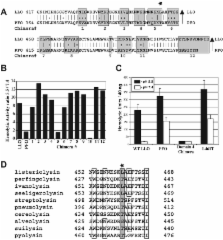
          Listeria monocytogenes is a facultative intracellular bacterial pathogen that escapes from a phagosome and grows in the host cell cytosol. The pore-forming cholesterol-dependent cytolysin, listeriolysin O (LLO), mediates bacterial escape from vesicles and is ∼10-fold more active at an acidic than neutral pH. By swapping dissimilar residues from a pH-insensitive orthologue, perfringolysin O (PFO), we identified leucine 461 as unique to pathogenic Listeria and responsible for the acidic pH optimum of LLO. Conversion of leucine 461 to the threonine present in PFO increased the hemolytic activity of LLO almost 10-fold at a neutral pH. L. monocytogenes synthesizing LLO L461T, expressed from its endogenous site on the bacterial chromosome, resulted in a 100-fold virulence defect in the mouse listeriosis model. These bacteria escaped from acidic phagosomes and initially grew normally in cells and spread cell to cell, but prematurely permeabilized the host membrane and killed the cell. These data show that the acidic pH optimum of LLO results from an adaptive mutation that acts to limit cytolytic activity to acidic vesicles and prevent damage in the host cytosol, a strategy also used by host cells to compartmentalize lysosomal hydrolases.

          Related collections

          Most cited references26

          • Record: found
          • Abstract: found
          • Article: not found

          RADIOAUTOGRAPHIC STUDIES OF CHOLINE INCORPORATION INTO PERIPHERAL NERVE MYELIN

          This radioautographic study was designed to localize the cytological sites involved in the incorporation of a lipid precursor into the myelin and the myelin-related cell of the peripheral nervous system. Both myelinating and fully myelinated cultures of rat dorsal root ganglia were exposed to a 30-min pulse of tritiated choline and either fixed immediately or allowed 6 or 48 hr of chase incubation before fixation. After Epon embedding, light and electron microscopic radioautograms were prepared with Ilford L-4 emulsion. Analysis of the pattern of choline incorporation into myelinating cultures indicated that radioactivity appeared all along the length of the internode, without there being a preferential site of initial incorporation. Light microscopic radioautograms of cultures at varying states of maturity were compared in order to determine the relative degree of myelin labeling. This analysis indicated that the myelin-Schwann cell unit in the fully myelinated cultures incorporated choline as actively as did this unit in the myelinating cultures. Because of technical difficulties, it was not possible to determine the precise localization of the incorporated radioactivity within the compact myelin. These data are related to recent biochemical studies indicating that the mature myelin of the central nervous system does incorporate a significant amount of lipid precursor under the appropriate experimental conditions. These observations support the concept that a significant amount of myelin-related metabolic activity occurs in mature tissue; this activity is considered part of an essential and continuous process of myelin maintenance and repair.
            Bookmark
            • Record: found
            • Abstract: found
            • Article: not found

            Gene splicing by overlap extension: tailor-made genes using the polymerase chain reaction.

            Gene Splicing by Overlap Extension or "gene SOEing" is a PCR-based method of recombining DNA sequences without reliance on restriction sites and of directly generating mutated DNA fragments in vitro. By modifying the sequences incorporated into the 5'-ends of the primers, any pair of polymerase chain reaction products can be made to share a common sequence at one end. Under polymerase chain reaction conditions, the common sequence allows strands from two different fragments to hybridize to one another, forming an overlap. Extension of this overlap by DNA polymerase yields a recombinant molecule. This powerful and technically simple approach offers many advantages over conventional approaches for manipulating gene sequences.
              Bookmark
              • Record: found
              • Abstract: found
              • Article: not found

              Actin filaments and the growth, movement, and spread of the intracellular bacterial parasite, Listeria monocytogenes

              Listeria monocytogenes was used as a model intracellular parasite to study stages in the entry, growth, movement, and spread of bacteria in a macrophage cell line. The first step in infection is phagocytosis of the Listeria, followed by the dissolution of the membrane surrounding the phagosome presumably mediated by hemolysin secreted by Listeria as nonhemolytic mutants remain in intact vacuoles. Within 2 h after infection, each now cytoplasmic Listeria becomes encapsulated by actin filaments, identified as such by decoration of the actin filaments with subfragment 1 of myosin. These filaments are very short. The Listeria grow and divide and the actin filaments rearrange to form a long tail (often 5 microns in length) extending from only one end of the bacterium, a "comet's tail," in which the actin filaments appear randomly oriented. The Listeria "comet" moves to the cell surface with its tail oriented towards the cell center and becomes incorporated into a cell extension with the Listeria at the tip of the process and its tail trailing into the cytoplasm behind it. This extension contacts a neighboring macrophage that phagocytoses the extension of the first macrophage. Thus, within the cytoplasm of the second macrophage is a Listeria with its actin tail surrounded by a membrane that in turn is surrounded by the phagosome membrane of the new host. Both these membranes are then solubilized by the Listeria and the cycle is repeated. Thus, once inside a host cell, the infecting Listeria and their progeny can spread from cell to cell by remaining intracellular and thus bypass the humoral immune system of the organism. To establish if actin filaments are essential for the spread of Listeria from cell to cell, we treated infected macrophages with cytochalasin D. The Listeria not only failed to spread, but most were found deep within the cytoplasm, rather than near the periphery of the cell. Thin sections revealed that the net of actin filaments is not formed nor is a "comet" tail produced.
                Bookmark

                Author and article information

                Journal
                J Cell Biol
                The Journal of Cell Biology
                The Rockefeller University Press
                0021-9525
                1540-8140
                18 March 2002
                : 156
                : 6
                : 1029-1038
                Affiliations
                [1 ]Department of Molecular and Cell Biology, University of California, Berkeley, CA 94720
                [2 ]The School of Public Health, University of California, Berkeley, CA 94720
                [3 ]Department of Microbiology and Immunology, University of Michigan Medical School, Ann Arbor, MI 48109
                Author notes

                Address correspondence to Daniel A. Portnoy, Department of Molecular and Cell Biology, 401 Barker Hall, University of California, Berkeley, CA 94703-3202. Tel.: (510) 643-3925. Fax: (510) 643-6791. E-mail: portnoy@ 123456uclink4.berkeley.edu

                Article
                0201081
                10.1083/jcb.200201081
                2173464
                11901168
                5a1abab5-5f08-45f2-a467-1fad36f627eb
                Copyright © 2002, The Rockefeller University Press
                History
                : 18 January 2002
                : 4 February 2002
                : 4 February 2002
                Categories
                Article

                Cell biology
                cytolysins; phagosome; virulence; molecular evolution; macrophages
                Cell biology
                cytolysins; phagosome; virulence; molecular evolution; macrophages

                Comments

                Comment on this article