9
views
0
recommends
+1 Recommend
0 collections
    0
    shares
      • Record: found
      • Abstract: found
      • Article: found
      Is Open Access

      Imaging in the diagnosis of ulnar nerve pathologies—a neoteric approach

      review-article

      Read this article at
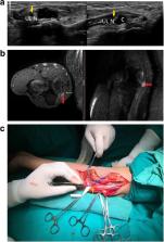
      Bookmark
          There is no author summary for this article yet. Authors can add summaries to their articles on ScienceOpen to make them more accessible to a non-specialist audience.

          Abstract

          The ulnar nerve is a branch of the C8 and T1 nerve roots and arises from the medial cord of the brachial plexus. It supplies the intrinsic muscles of the hand and assists the median nerve in functioning of the flexors. Also known as the musician’s nerve, it is the second most common nerve involved in compressive neuropathy following the median nerve. Common sites of entrapment include cubital tunnel at the elbow, the ulnar groove in the humerus and the Guyon’s canal at the wrist. Patients present with altered sensation in the ulnar fourth and the fifth digit and the medial side of arm with loss of function of intrinsic muscles of the hand, the flexor carpi ulnaris and ulnar fibres of flexor digitorum superficialis in more severe cases. Diagnosis relies on clinical examination, electrodiagnostic studies and imaging findings. Plain radiographs are used to identify fracture sites, callus, or tumours as cause of compression. Technological advances in ultrasonography have allowed direct visualisation of the involved nerve with assessment of exact site, extent and type of injury. It yields unmatched information about anatomical details of the nerve. MR imaging adds to soft tissue details and helps in characterising the lesion. This pictorial review aims to illustrate a wide spectrum of causes of ulnar neuropathies as seen on ultrasound and MRI and emphasises upon the importance of imaging modalities in the diagnosis of neuropathies.

          Related collections

          Most cited references42

          • Record: found
          • Abstract: found
          • Article: not found

          Peripheral nerves of the extremities: imaging with US.

          B. Fornage (1988)
          High-resolution real-time ultrasonography (US) was used to evaluate peripheral nerves of the extremities in healthy subjects and in 11 patients with a mass developed from a peripheral nerve. The normal median and ulnar nerves in the upper extremity and the normal sciatic and external popliteal nerves in the lower extremity were seen, all having an echogenic fibrillar echotexture. Pathologic findings included nine cases of benign tumor (four schwannomas, three neurofibromas, two traumatic neuromas), one of neurilemmitis, and one of tuberculoid leprosy. All lesions were hypoechoic. Three of the four schwannomas had well-defined contours, and two were associated with a typical distal sound enhancement. Neurofibromas and traumatic neuromas were less sharply delineated. Inflammatory conditions were characterized by a hypoechoic, thickened nerve. US was effective in imaging nerve masses in the extremities, and large normal nerves can now be demonstrated with high-resolution US.
            Bookmark
            • Record: found
            • Abstract: found
            • Article: not found

            Ultrasound in the diagnosis of ulnar neuropathy at the cubital tunnel.

            Ulnar neuropathy at the cubital tunnel (UCT) is diagnosed on the basis of history, physical examination, and nerve conduction studies (NCSs); however, the wide spectrum of findings often makes the diagnosis difficult. The purpose of this study was to document the ultrasonographic differences in ulnar nerve size between patients with UCT and control subjects, and to correlate those differences with clinical examination findings and NCS abnormalities, thereby testing the validity of ultrasound (US) as an additional adjunct diagnostic modality for UCT. Fifteen elbows in 14 patients with symptoms, clinical examination, and NCS findings consistent with UCT had US of the ulnar nerve. Patients were excluded if they had a history of polyneuropathy, acute trauma involving the upper extremity, previous trauma in the region of the elbow (including previous surgery), or brachial plexus injury. The control group consisted of 60 elbows from 30 normal volunteers that also had US. Maximal cross-sectional areas (CSAs) were measured and compared for the 2 groups and a correlation analysis was performed between nerve size and NCS findings. The average CSA of the ulnar nerve was 0.065 cm(2) in the control group, whereas in the UCT group it was 0.19 cm(2), indicating a significant statistical difference in ulnar nerve size between the 2 groups. The Pearson correlation coefficient between motor nerve conduction velocity of the ulnar nerve and the CSA was 0.80. High-resolution US is a noninvasive, safe, and reliable modality for imaging the ulnar nerve at the elbow and it may provide a valuable adjunct to NCS in the diagnosis of UCT.
              Bookmark
              • Record: found
              • Abstract: found
              • Article: not found

              Nerve entrapment syndromes of the elbow, forearm, and wrist.

              Nerve entrapment syndromes usually have typical clinical presentations and findings on physical examination. Imaging can be used to evaluate a structural cause of the entrapment, such as a mass or enlarged muscle or to show secondary findings that confirm the diagnosis, such as nerve swelling or muscle edema or atrophy. This article will review entrapment syndromes of the elbow, forearm, and wrist.
                Bookmark

                Author and article information

                Contributors
                9510148944 , 8789767041 , a.agarwal.1992@gmail.com
                Journal
                Insights Imaging
                Insights Imaging
                Insights into Imaging
                Springer Berlin Heidelberg (Berlin/Heidelberg )
                1869-4101
                20 March 2019
                20 March 2019
                December 2019
                : 10
                : 37
                Affiliations
                [1 ]ISNI 0000 0004 1767 3615, GRID grid.416077.3, Department of Radiodiagnosis, , SMS Medical College, ; Jaipur, Rajasthan India
                [2 ]ISNI 0000 0004 1767 3615, GRID grid.416077.3, Department of Orthopaedics, , SMS Medical College, ; Jaipur, Rajasthan India
                [3 ]A 235, Shivanand Marg, Malviya Nagar, Jaipur, India
                Article
                714
                10.1186/s13244-019-0714-x
                6426899
                30895491
                580995d7-b71e-4a61-bb09-a4207ebf8f26
                © The Author(s). 2019

                Open AccessThis article is distributed under the terms of the Creative Commons Attribution 4.0 International License ( http://creativecommons.org/licenses/by/4.0/), which permits unrestricted use, distribution, and reproduction in any medium, provided you give appropriate credit to the original author(s) and the source, provide a link to the Creative Commons license, and indicate if changes were made.

                History
                : 16 November 2018
                : 4 February 2019
                Categories
                Pictorial Review
                Custom metadata
                © The Author(s) 2019

                Radiology & Imaging
                imaging in the ulnar mononeuropathy,high-resolution ultrasound of ulnar neuropathy,mr ulnar neuropathy

                Comments

                Comment on this article