There is no author summary for this article yet. Authors can add summaries to their articles on ScienceOpen to make them more accessible to a non-specialist audience.
Abstract
Leprosy is an infectious disease caused by Mycobacterium leprae that affects the skin
and nerves, presenting a singular clinical picture. Across the leprosy spectrum, lepromatous
leprosy (LL) exhibits a classical hallmark: the presence of a collection of M. leprae-infected
foamy macrophages/Schwann cells characterised by their high lipid content. The significance
of this foamy aspect in mycobacterial infections has garnered renewed attention in
leprosy due to the recent observation that the foamy aspect represents cells enriched
in lipid droplets (LD) (also known as lipid bodies). Here, we discuss the contemporary
view of LD as highly regulated organelles with key functions in M. leprae persistence
in the LL end of the spectrum. The modern methods of studying this ancient disease
have contributed to recent findings that describe M. leprae-triggered LD biogenesis
and recruitment as effective mycobacterial intracellular strategies for acquiring
lipids, sheltering and/or dampening the immune response and favouring bacterial survival,
likely representing a fundamental aspect of M. leprae pathogenesis. The multifaceted
functions attributed to the LD in leprosy may contribute to the development of new
strategies for adjunctive anti-leprosy therapies.
The lipid droplet (LD) is an organelle that is used for the storage of neutral lipids. It dynamically moves through the cytoplasm, interacting with other organelles, including the endoplasmic reticulum (ER). These interactions are thought to facilitate the transport of lipids and proteins to other organelles. The hepatitis C virus (HCV) is a causative agent of chronic liver diseases. HCV capsid protein (Core) associates with the LD, envelope proteins E1 and E2 reside in the ER lumen, and the viral replicase is assumed to localize on ER-derived membranes. How and where HCV particles are assembled, however, is poorly understood. Here, we show that the LD is involved in the production of infectious virus particles. We demonstrate that Core recruits nonstructural (NS) proteins and replication complexes to LD-associated membranes, and that this recruitment is critical for producing infectious viruses. Furthermore, virus particles were observed in close proximity to LDs, indicating that some steps of virus assembly take place around LDs. This study reveals a novel function of LDs in the assembly of infectious HCV and provides a new perspective on how viruses usurp cellular functions.
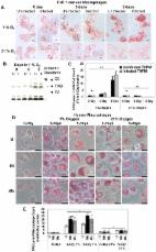
Introduction One-third of the world population is latently infected with Mycobacterium tuberculosis (Mtb) and this vast reservoir is expected to contribute towards an increasing incidence of tuberculosis (TB) disease. The World Health Organization estimated recently that there were 11 million prevalent cases of the disease and 1.8 million deaths annually due to TB, including 0.5 million deaths in HIV-positive patients [1]. Mtb, the causative agent, is inhaled as an aerosol and enters the lung where it infects the alveolar macrophages and eludes host defenses. The primary immune response of the host controls bacillary multiplication and causes the pathogen to enter a state of dormancy and become phenotypically antibiotic tolerant leading to latent TB [2], [3], [4]. As a result of the host immune response, the pathogen is contained within the granuloma which is made up of infected macrophages surrounded by foamy lipid-loaded macrophages, mononuclear phagocytes and lymphocytes enclosed within a fibrous layer of endothelial cells [5], [6], [7]. Mtb can persist inside the host for decades until the host immune system is weakened and then reactivates to cause active disease [3]. It was established several decades ago that Mtb inside the host uses fatty acids as the major source of energy [8]. Isocitrate lyase (icl), which has been known to be a key enzyme of the glyoxylate cycle used by organisms that live on fatty acids [9], was shown to be vital for the pathogen's persistence inside the host demonstrating the critical role of fatty acids as an energy source for Mtb [10]. Based on the observation that fatty acids are normally stored as triacylglycerol (TAG) in the adipose tissues of mammals, seed oils of plants and as lipid inclusion bodies in prokaryotes for use as energy source during and after dormancy/ hibernation, TAG was postulated to be the storage form of energy for latent Mtb [11]. Intracellular lipid inclusion bodies were initially observed in Mtb more than six decades ago and were more recently detected in mycobacteria isolated from the sputum of TB patients [12], [13]. We showed that TAG accumulation is a critical event of Mtb dormancy and reported the discovery of triacylglycerol synthase 1 (tgs1) as the primary contributor to TAG synthesis within the pathogen and that the deletion of tgs1 led to a nearly complete loss in TAG accumulation by Mtb under in vitro dormancy-inducing conditions [11], [14], [15]. Recent observations from other groups have shown that the tgs1 gene is upregulated and TAG accumulates in dormant Mtb found in the sputum of TB patients and in the widespread, multi-drug resistant W/Beijing strain of Mtb [16], [17]. The source of fatty acids for synthesis of the TAG that accumulates as lipid droplets in the pathogen remains unknown. In humans with untreated pulmonary TB, caseous granulomas in the lungs were shown to contain lipid-loaded foamy macrophages which harbored acid-fast bacilli [7]. Such lipid-loaded macrophages which are found inside the hypoxic environment of the tuberculous granuloma contain abundant stores of TAG and are thought to provide a lipid-rich microenvironment for Mtb [5], [6]. Human macrophages cultured under hypoxia (1% O2) accumulate TAG in lipid droplets [18]. Mtb-infected human alveolar macrophages are most likely enclosed in a hypoxic environment within the granuloma where the pathogen becomes dormant. It was shown recently that tuberculous granulomas in guinea pigs, rabbits and non-human primates were hypoxic [19]. It is well recognized that nonpulmonary tissue oxygen concentrations within the human body are far below the oxygen concentration in ambient room air and the typical oxygen level in standard in vitro cell cultures is much higher than that encountered by macrophages inside the human body [20], [21]. Furthermore, the oxygen concentration in the phagosome of activated macrophages was shown to be lower than the extracellular oxygen concentration [22]. Dissemination of Mtb to distal sites such as the adipose tissue may also provide a TAG-enriched host environment for Mtb to go into dormancy [23]. We postulate that Mtb inside lipid-loaded macrophages might import fatty acids derived from host TAG to accumulate TAG inside the bacterial cell and provide evidence to support this hypothesis. We infected human peripheral blood mononuclear cell (PBMC)-derived macrophages and THP-1 derived macrophages (THPM) with Mtb and incubated them under hypoxia (1% O2) in order to mimic the microenvironment within the human lung granuloma. We demonstrate that the macrophages accumulate lipid droplets under hypoxia. Using single and double isotope labeling methods to metabolically label the host TAG, we determined that Mtb imports fatty acids released from host TAG to accumulate TAG within the bacterial cell. Host fatty acids were incorporated intact into Mtb TAG. We also show that host TAG that was metabolically labeled with a fluorescent fatty acid was imported by Mtb and accumulated as fluorescent lipid droplets within the bacterial cell. Deletion of tgs1 resulted in a drastic decrease in radiolabeled and fluorescent TAG accumulation within Mtb inside THPM thereby revealing that synthesis of TAG within the pathogen from fatty acids released from host TAG constitutes the major pathway of TAG accumulation by Mtb inside the host. We demonstrate that Mtb cells within lipid-loaded macrophages accumulate lipid droplets containing TAG, lose acid-fast staining and become phenotypically resistant to the two frontline antimycobacterial drugs, rifampicin (Rif) and isoniazid (INH), all of which are thought to be indicative of the dormant state of the pathogen [2], [11], [15], [24], [25]. Taqman real-time PCR analysis of gene transcripts of Mtb recovered from lipid-loaded macrophages revealed that genes thought to be involved in dormancy and lipid metabolism were upregulated within the pathogen. Results Macrophages accumulate triacylglycerol in lipid droplets under hypoxia Human alveolar macrophages, in which Mtb multiplies, probably reach a hypoxic environment within the granuloma, in which the pathogen goes into a latent state. Such macrophages are likely to be lipid-loaded as a consequence of hypoxia and Mtb infection, both of which have been reported to induce lipid accumulation in macrophages in vitro [6], [18], [26]. It is well known that nonpulmonary tissue oxygen concentrations within the human body are much lower than the oxygen level in ambient air and that caseous granulomas in rabbits are hypoxic [19], [21]. In order to mimic the hypoxic microenvironment within the granuloma, we infected human PBMC-derived macrophages and THPM with low numbers of Mtb (MOI 0.1 to 5) and incubated them under 1% O2, 5% CO2. About 3% of the host cells were infected at MOI 0.1 as determined by the CFUs recovered from the infected host cells after 4 h infection. Oil Red O-staining lipid bodies increased upto 5 days in Mtb-infected macrophages as well as uninfected macrophages incubated under 1% O2 (Figure 1A, D). In contrast, lipid bodies increased moderately in macrophages incubated under 21% O2 (Figure 1A, D). TAG was the major lipid that accumulated in THPM lipid droplets under hypoxia and maximal levels were reached by day 5 (Figure 1B and C). Longer incubations resulted in greater loss of THPM from the adhered monolayer (data not shown). TAG accumulation in lipid bodies was also strongly induced under hypoxia in human PBMC-derived macrophages (Figure 1 D, E). Lipid droplets containing TAG increased greatly in size and number with time of culturing under hypoxia but only moderately under normoxia, and when normalized to viable macrophage cell counts, it was observed that TAG levels in hypoxic macrophages were much higher than that in normoxic macrophages. There were considerable differences in lipid body formation between macrophages in the same population. Since the photomicrographs showing selected fields of Oil Red O-stained macrophages do not adequately represent the TAG levels in the whole macrophage population, we relied on the analysis by thin-layer chromatography (TLC) of the TAG levels in the lipid extracts from the total population. 10.1371/journal.ppat.1002093.g001 Figure 1 Macrophages under hypoxia accumulate Oil Red O staining lipid droplets containing triacylglycerol. A, Oil Red-O stained lipid droplets increase in THPM under 1 % O2 independent of Mtb infection but do not increase in THPM incubated under 21% O2. THPM were infected with Mtb at an MOI of 0.1 and incubated under hypoxia or normoxia as described in Methods. At each time-point, trypsinized THPM were fixed with 4% paraformaldehyde and stained with Oil Red-O. Uninfected THPM were incubated under identical conditions. Scale bar, 10 µm. B, Silica-TLC of lipid extracts from uninfected (U) and infected (I) hypoxic THPM. The lipid extracts were resolved on silica-TLC and visualized by dichromate-sulfuric acid charring as described in Methods. Relative migrations of authentic standard cholesteryl ester (CE), TAG and fatty acid (FA) are shown. C, TAG levels in THPM increase under hypoxia but not under normoxia. TAG band intensities on TLC plates were normalized to the respective viable THPM cell counts and represented as fold of 0-day levels. 0-day levels were assigned an arbitrary value of 1. Data is represented as average ± SD from a representative experiment (n = 3). *, Statistically insignificant difference (p>0.05) between uninfected and infected THPM; **, statistically significant difference (p 0.05) between 1% vs 21% incubations;. C, Mtb replication inside hypoxic THPM is severely curtailed in contrast to normoxic THPM. THPM were infected at an MOI of 0.1 and incubated under 1% O2 or 21% O2. At 0, 3, 5-days, Mtb CFUs were determined by agar plating. Mtb CFUs were normalized to THPM numbers. Data from triplicate measurements presented as average ± SD (n = 3); *, statistically significant differences (p<0.05) between 1% O2 vs. 21% O2 incubations. Viability of PBMC-derived macrophages (infected and uninfected) in the adhered monolayer was about 97% at 3 days and 92 % at 5 days under 1% O2 at both MOIs. At 3 and 5 days under normoxia, about 95% of the adhered human macrophages (infected and uninfected) were viable. The total viable cell counts (by Trypan Blue dye exclusion method) of adhered hypoxic human macrophages at 3 and 5 days were about 30% of the 0-day count of 5×10⁵ macrophages per well of a 12-well plate and the respective counts for normoxic samples were about 45% of the 0-day count. We determined the rate of Mtb multiplication within macrophages under 21% O2 and 1% O2. After normalization to the respective THPM cell counts, Mtb CFUs inside THPM under 1% O2 at day 5, increased to about 5-fold of 0-day values. In contrast, Mtb CFUs inside normoxic THPM increased to about 30-fold of 0-day values by day 5 (Figure 6C). Mtb CFUs in the extra-cellular medium were much lower than those inside adhered THPM monolayer (data not shown). Mtb replication within PBMC-derived macrophages under hypoxia was even more restricted than that inside hypoxic THPM. At day 5 under hypoxia, Mtb CFUs in PBMC-derived macrophages normalized to macrophage cell count was about 3-fold of 0-day values. In contrast, Mtb CFUs increased to about 34-fold of 0-day values at day 5 inside human PBMC-derived macrophages incubated under normoxia. Mtb inside lipid-loaded macrophages develops antibiotic resistance If the microenvironment inside hypoxic lipid-loaded macrophages mimics what happens in the hypoxic environment of the granuloma, we might expect Mtb within such macrophages to develop phenotypic drug resistance which is a key indicator of dormancy [2], [4], [15]. To test for this possibility, we examined whether such phenotypic tolerance may be developed by Mtb within THPM and inside human PBMC-derived macrophages under 1% O2. At 0, 3 and 5 days after infection, Mtb cells inside macrophages were exposed to antibiotic for 2 additional days, under the same conditions, prior to lysis of the host cells and recovery of the bacilli. The antibiotic resistance, as a percentage of untreated control incubated for the same time-period under the same oxygen concentration, was determined by CFU determination after agar plating. As shown in Table 3, we found that phenotypic tolerance of Rif and INH of Mtb recovered from hypoxic THPM increased with time and reached maximal levels by 5 days under 1% O2 when about 8% of the total Mtb population was resistant to 5 µg/ml Rif and about 49% was resistant to 0.8 µg/ml INH. Further incubation (upto 16 days) in 1% O2 decreased the percentage of antibiotic-resistant Mtb (data not shown). In contrast, Mtb inside normoxic THPM did not develop phenotypic tolerance of the antibiotics (data not shown). Mtb inside human PBMC-derived macrophages incubated under hypoxia also developed phenotypic tolerance to Rif and INH, as observed in THPM (Table 3). Phenotypic resistance to Rif and INH increased to 18% and 43% respectively at day 7 inside hypoxic PBMC-derived macrophages. In contrast, Mtb inside normoxic human macrophages showed much lower phenotypic tolerance to Rif (4%) and negligible phenotypic tolerance (0.5%) to INH at day 7 under normoxia. Log-phase Mtb cultures used for infection and Mtb recovered from macrophages after 4 h infection and treated in vitro with antibiotics under normoxia for 2 days showed no resistance to Rif and INH. Thus, Mtb developed phenotypic drug tolerance in hypoxic THPM as well as in hypoxic human PBMC-derived macrophages. 10.1371/journal.ppat.1002093.t003 Table 3 Mtb within hypoxic lipid-loaded macrophages develops phenotypic tolerance to antibiotics. THPM or human PBMC-derived macophages infected with Mtb at an MOI of 0.1 were incubated in 1 % O2, 5 % CO2 or 21 % O2, 5 % CO2. At 0, 3 and 5 days after infection, the infected host cells were treated with antibiotics at the indicated concentrations for 2 more days under the same conditions prior to lysis of the host cells. Mtb recovered from antibiotic-treated macrophages were analyzed for antibiotic resistance and compared to Mtb recovered from macrophages unexposed to antibiotics, by CFU plating. Mtb inside hypoxic lipid-loaded macrophages accumulates neutral lipid bodies and loses acid fastness It has been established previously that dormant Mtb loses acid-fast staining and accumulates Nile Red-staining lipid droplets [13], [15], [16], [25]. In order to determine whether such a phenotype is developed by Mtb inside hypoxic lipid-loaded macrophages, Mtb cells recovered from human PBMC-derived macrophages after 0, 3 and 5 days in 1% O2 were stained with Auramine-O and Nile Red. We observed that, in addition to the bacilli that stained with either stain, there was a subset of bacilli in the total population that retained both stains. The fraction of the Mtb population that stained with the green acid-fast stain (Auramine-O) decreased from about 86% at 0-day to about 40% at day 5. In contrast, Mtb cells that stained red with the lipid stain (Nile Red) increased with time from about 35% at 0-day to about 81% at 5-day inside hypoxic human macrophages (Figure 7A–D). Thus, by day 5 inside hypoxic macrophages, the fraction of acid-fast staining bacilli in the Mtb population decreased to half the level of the 0-day control, while the fraction that stained with Nile Red increased more than two-fold. Moreover, at day 5 inside hypoxic macrophages, Mtb cells were markedly elongated in shape when compared to the 0-day controls. In order to stain Mtb inside intact host cells, infected THPM after 5 days in 1% O2 were fixed with 4 % paraformaldehyde and stained with Auramine-O followed by Nile Red. Mtb cells inside such intact THPM showed loss of acid-fastness and accumulation of Nile Red staining lipid droplets similar to the Mtb cells that were recovered from the macrophages before staining (Figure 7E–G). 10.1371/journal.ppat.1002093.g007 Figure 7 Mtb inside hypoxic macrophages loses acid fastness and accumulates lipid droplets. A–C, Decrease in green, Auramine-O staining, acid-fast positive Mtb and increase in Nile Red staining, neutral lipid-containing Mtb population recovered from hypoxic human macrophages with time. Human macrophages were infected at MOI 0.1 and were incubated at 1% O2, 5% CO2 at 37°C. Mtb cells were recovered from human macrophages after 4 h infection (A), at 3 days (B) and 5 days (C) and stained with Auramine-O and Nile Red; D, Quantitation of acid-fast and neutral lipid staining Mtb recovered from hypoxic human macrophages (shown in A–C) indicates a decrease in acid-fastness and increase in lipid droplet staining with time. About 250 Mtb cells from multiple microscopic fields were counted for enumerating green and red cells. E–G, Mtb within intact hypoxic THPM at day 5 showing loss of acid-fastness (green Auramine-O stain negative) and accumulation of lipid bodies (Nile Red stain positive) by confocal laser scanning microscopy. Infected THPM were subjected to 1% O2, 5% CO2 for 5 days at 37°C. Sequential laser scanning was done for Auramine-O (E) and for Nile Red (F); G, Merged projection of E and F. Genes associated with dormancy and lipid metabolism are upregulated in Mtb within THPM We examined the changes in transcript levels of selected Mtb genes that have been shown to be upregulated in a variety of in vitro and in vivo experimental models that mimicked dormancy [27]. The gene for isocitrate lyase (icl) was induced (Figure 8), consistent with the idea that the pathogen in THPM utilizes fatty acids as the energy source. Induction of dormancy- and stress-responsive genes, dosR (Rv3133c) and hspX (Rv2031c), implicates the attainment of the dormant state by Mtb inside hypoxic, lipid-loaded THPM. In our hypoxic THPM model, tgs1 (Rv3130c), Rv3088 (tgs4), Rv1760, Rv3371 and Rv3087 (data not shown for this gene) were found to be highly up-regulated at 72 h after infection. It is noteworthy that lipY, that was previously reported to be involved in TAG mobilization [28], was highly induced. Induction of other lipase and cutinase-like genes suggests their possible involvement in the hydrolysis of host lipids. The fatty acyl-coenzyme A reductase (fcr) genes Rv3391 and Rv1543, that are involved in wax ester biosynthesis ([15], unpublished results) were also upregulated. 10.1371/journal.ppat.1002093.g008 Figure 8 Dormancy and lipid metabolism genes are upregulated in Mtb recovered from lipid-loaded macrophages. TaqMan real-time PCR was used to measure the transcript levels of Mtb genes reported to be highly upregulated in a meta-analysis of Mtb microarray data from experimental models that mimicked dormancy. Mtb was recovered from lipid-loaded host cells at 72 h after incubation under hypoxia (1% O2; 5% CO2). Total RNA was reverse transcribed, the resulting cDNA was pre-amplified by multiplex-PCR with multiple Mtb gene-specific primers and the pre-amplified product was used in quantitative (q) PCR. Data was analyzed by ‘GenEx’ qPCR data analysis software (MultiD Analyses AB, Sweden) and gene transcript level was expressed as fold change in log2 scale relative to the sample from 18 h time point following normalization with 16S-rRNA as the reference gene. Average ± standard deviation from three replicates shown (n = 3); p<0.05, 18 h vs 72 h. lip, lipase, tgs, triacylglycerol synthase, cut, cutinase, fcr, fatty acyl-CoA reductase, icl, isocytrate lyase, dosR, dormancy response regulator, hsp, heat shock protein. The number prefixes are gene locus tag (Rv) numbers for respective Mtb genes. Discussion Mtb can persist for decades inside the human body in the dormant state and reactivate when the host's immune system weakens [4]. HIV infection increases the risk of reactivation leading to the deadly synergy between AIDS and TB [3], [29], [30]. Currently, there is no drug that can kill latent TB and the development of such antibiotics is critical to the cure and eradication of the disease [2], [31]. Novel drugs that target dormancy-specific metabolic pathways may enable the treatment of patients with multi- and extremely-drug resistant Mtb and drastically shorten the currently used, very long-term treatment period to cure TB. Understanding of dormancy-specific processes and a model system to test for inhibition of such processes are required to discover such drugs. The pathogen is likely to go into a dormant state within macrophages that are in the hypoxic environment of the granuloma [15], [32], [33]. Such macrophages might be loaded with TAG-containing lipid bodies [6], [7], [18]. Since one of our objectives was to develop an in vitro model that mimics the in vivo situation and is suitable for high-throughput screening, we used THPM as host cells in order to avoid the well known donor-to-donor variations in primary human macrophages and the technical difficulties involved in obtaining large, homogenous populations of alveolar macrophages for experimental purposes. We validated our results obtained with hypoxic THPM by demonstrating similar observations in human macrophages which were derived from mononuclear cells isolated from the peripheral blood of healthy volunteers and subjected to hypoxia. THPM, which are capable of lipid accumulation, were reported to faithfully model the apoptotic response of human alveolar macrophages in response to Mtb infection [34], [35]. Furthermore, the antimycobacterial activity of INH in THPM was similar to that in human monocyte-derived macrophages [36]. The assumption that Mtb-infected human alveolar macrophages most likely reach a hypoxic environment within the granuloma serves as the basis for the well-studied in vitro hypoxic model of Mtb dormancy [33]. Moreover, oxygen concentrations in healthy tissue within the human body are thought to range between 5 to 71 Torr and are well below the oxygen concentration of 157 Torr in ambient room air [20], [21]. The oxygen tension in caseous granulomas of rabbits was measured to be approximately 2 Torr (∼0.3 % O2) [19]. Hypoxic, lipid-loaded macrophages may provide a lipid-rich sanctuary for Mtb during its dormancy. The killing of Mtb by macrophages inside the hypoxic regions of the granuloma is likely to be severely inhibited since superoxide and NO production by macrophages are greatly diminished by hypoxia [21], [37]. Furthermore, electron paramagnetic resonance-based measurements have shown that oxygen concentration in the intraphagosomal compartment was significantly lower than the extracellular environment [22]. However, macrophages infected in vitro with Mtb are currently incubated in normoxic environments where the oxygen level is far higher than that encountered by Mtb-infected macrophages inside the human lung granuloma. Consequently, Mtb inside those macrophages are not subjected to the hypoxic stress encountered inside the granuloma and do not develop phenotypic tolerance of antibiotics such as Rif and INH [36], [38] which is a key indicator of dormancy [2], [4], [15]. In order to mimic the hypoxic micro-environment within the granuloma, we infected macrophages with Mtb and incubated them in a 1% O2, 5% CO2 environment. Under such conditions, infected and uninfected macrophages accumulated Oil Red O-staining lipid droplets containing TAG. The replication of Mtb within such hypoxic lipid-loaded macrophages was greatly inhibited suggesting that a subset of the Mtb inside macrophages incubated under hypoxia may be entering a non-replicating state. Interestingly, hypoxia (1% O2) was recently shown to prolong the survival of human macrophages and the cells were reported to be adopting a glycolytic metabolism under the hypoxic conditions [39]. We postulate that host lipids may be hydrolyzed by Mtb lipases and the released fatty acids may be imported and re-esterified into Mtb TAG by the action of Mtb tgs gene products. The deletion of Mtb tgs1 gene, which encodes the major TAG biosynthetic enzyme of Mtb [11], [14], resulted in a severe decrease of radiolabeled TAG accumulation by Mtb inside lipid-loaded THPM. Mtb inside lipid-loaded macrophages utilized host TAG that had been metabolically labeled with the fluorescent fatty acid BODIPY 558/568 C12 to accumulate fluorescent lipid droplets. Analysis of deconvoluted, Z-stacked fluorescence microscope images of Mtb recovered from fluorescent fatty acid-labeled THPM confirmed that the fluorescent lipid droplets are indeed inside the bacterial cell. Deletion of tgs1 drastically reduced fluorescent lipid droplet accumulation and supported the finding from the radiolabeling experiments suggesting that TGS1 is a major contributor to TAG synthesis within Mtb. TGS1, which has very recently been shown to be associated with lipid droplets in the mycobacterial cell along with TGS2 [40], is most likely involved in Mtb lipid droplet synthesis. Since TAG accumulation in the tgs1 mutant was not totally abolished, the other Mtb tgs gene products might also be able to contribute to TAG synthesis within Mtb inside the host in the absence of tgs1. In order to assess whether Mtb inside THPM imported intact host TAG or hydrolyzed the host TAG and imported the fatty acids released, we metabolically labeled the TAG in THPM using dual-isotope labeled triolein. The glycerol backbone of the triolein was radiolabeled with 3H and the esterified fatty acids were labeled at the carboxyl end with 14C. By comparing the 3H:14C ratios of TAG isolated from Mtb recovered from such dual-isotope labeled THPM with that of host TAG, we were able to conclude that the main mechanism by which host lipids are used to accumulate TAG within the pathogen involves the use of fatty acids released from host TAG for resynthesis of TAG within Mtb. Thus, Mtb gene products that are involved in the import of host-derived fatty acids and synthesis of TAG within Mtb may play critical roles in the energy metabolism of dormant Mtb. We cannot, however, rule out the possibility that the import of intact TAG might also make a contribution to TAG accumulation by Mtb inside the host. To determine whether host TAG-derived fatty acids were incorporated intact into TAG in Mtb within THPM or whether degradation of host-derived fatty acids and resynthesis of fatty acids contributed to lipid accumulation in Mtb, we labeled THPM TAG with [9,10-3H, 1-14C] labeled oleic acid. We observed that host TAG-derived fatty acids were being incorporated intact into Mtb TAG. Furthermore, if acetate derived from the catabolism of host TAG-derived fatty acids was used in the synthesis of fatty acids within Mtb, TAG of Mtb recovered from THPM should contain C26 fatty acid, a characteristic product of the Mtb fatty acid synthase [41]. The fatty acid composition of unlabeled Mtb TAG was identical to host TAG and C26 fatty acid was not detected in the TAG of Mtb recovered from THPM. Both the dual-isotope labeling experiments and fatty acid composition analysis of Mtb TAG, indicate that fatty acids released from host TAG were incorporated intact into Mtb TAG. The TAG levels in Mtb recovered from radiolabeled human macrophages were lower than that in Mtb recovered from THPM (Figure 2A). Possibly, inside hypoxic human macrophages, Mtb replication and metabolism is restricted more severely than in hypoxic THPM resulting in lower TAG synthesis by Mtb. This possibility correlates with our observation that shows greater antibiotic tolerance by Mtb in hypoxic human macrophages than in hypoxic THPM (Table 3). The quantity of TAG inside Mtb does not correspond directly with macrophage TAG levels probably because the quantity of TAG in the host is several orders of magnitude higher than that in the pathogen. The accumulation of neutral lipids, loss of acid-fastness and development of phenotypic antibiotic tolerance by Mtb are thought to be key indicators of dormancy [2], [11], [15], [24], [25]. We observed that a subset of the Mtb population within hypoxic, lipid-loaded macrophages accumulated neutral lipid droplets and lost acid-fast staining indicating their dormant state. The loss of acid-fastness by a subpopulation of Mtb inside hypoxic macrophages supports our hypothesis that Mtb cells inside the lipid-loaded macrophages enter a dormant state. Mtb recovered from hypoxic, lipid-loaded macrophages showed phenotypic resistance to killing by Rif and INH. The natural heterogeneity of the Mtb population within macrophages probably prevents the entire population from displaying a uniform dormancy phenotype. This is one of the possible causes for only a subset of the Mtb becoming tolerant to antibiotics and accumulating storage lipids. The drastically slowed replication rate of Mtb inside macrophages under hypoxia probably causes the observed phenotypic antibiotic resistance. We do not have a clear understanding of the reasons for our observation of a smaller percentage of Mtb recovered from hypoxic macrophages showing resistance to Rif in comparison to INH. Possibly, among the non-replicating, INH-resistant Mtb population, a subset of the Mtb is metabolically inactive and thus displays Rif-resistance. The earlier findings by Peyron et al showing that only a subset (19%) of the bacilli were translocated into the lipid bodies of the foamy macrophages inside in vitro granulomas and exhibited intracellular lipophilic inclusions [6] could offer another reason for our observations which show a subset of Mtb becoming phenotypically drug tolerant. Alternatively, TAG accumulation and phenotypic antibiotic tolerance may be independent indicators of Mtb dormancy. Preliminary results from our ongoing studies assessing drug tolerance of Mtb wild-type and Mtb tgs1-deletion mutant in hypoxic, lipid-loaded THPM indicate that the loss of tgs1 causes a small, but detectable decrease in antibiotic tolerance suggesting that TAG accumulation and phenotypic drug tolerance may be independent indicators of dormancy (our unpublished observations). In our earlier findings with the in vitro multiple-stress model, TAG accumulation and drug tolerance appeared to be strongly correlated [15]. A major difference between the two in vitro dormancy models is that Mtb inside the lipid-loaded macrophages is exposed to a readily available supply of host TAG in the lipid bodies whereas in the multiple stress model Mtb was cultured in a nutritionally limited environment (10% Dubos medium). It appears that the two indicators of dormancy, TAG accumulation and drug tolerance, show strong correlation only when Mtb experiences nutritionally limiting conditions. We found that at day 5 after infection, the antibiotic resistance of Mtb recovered from hypoxic lipid-loaded macrophages reached maximal levels. In normoxic macrophages, Mtb did not develop drug tolerance to the high levels seen in hypoxic, lipid-loaded macrophages (Table 3) probably due to the high rate of multiplication (Figure 6C). This finding is consistent with earlier reports that showed a lack of antibiotic resistance in Mtb inside normoxic macrophages [36], [38]. Thus, unlike in the conventional macrophages incubated under normoxia (our results and [36], [38]), in the hypoxic, lipid-loaded macrophages, Mtb displays intracellular TAG accumulation, phenotypic drug tolerance and loss of acid-fastness - the three key indicators of dormancy. A recent report showed that human macrophages infected with Mycobacterium leprae secreted TLR2 and TLR6 and caused uninfected macrophages to become lipid-loaded in addition to the infected macrophages [42]. It would be interesting to examine in future studies whether such paracrine signaling mechanisms play similar roles in hypoxic macrophages. We examined the transcript levels of selected Mtb genes involved in lipid metabolism and known to be up-regulated in a meta-analysis of Mtb microarray data from in vitro and in vivo experimental models that mimicked dormancy [27]. Interestingly, the tgs, lip, cut and fcr genes that received high up-regulation scores in the meta-analysis [27], were also found to be significantly induced in our hypoxic lipid-loaded THPM model. Several tgs genes, including tgs1, were induced indicating their possible involvement in the storage of fatty acids derived from host lipids as TAG within the pathogen, consistent with our hypothesis. The other tgs genes induced in this system might be responsible for the finding that TAG accumulation was not totally abolished in the tgs1 mutant. We reported previously that the Mtb lipase (LIPY), which belongs to the hormone-sensitive lipase family, was capable of releasing fatty acids from TAG stored within the pathogen for utilization during starvation [28]. LIPY has subsequently been shown by others to be localized on the mycobacterial cell wall and plays a major role in the hydrolysis of TAG within Mtb [43], [44]. The upregulation of the lipY gene in Mtb recovered from THPM is consistent with the observation that the TAG that accumulates in the pathogen is generated within Mtb from fatty acids released from host TAG and suggests its possible involvement in releasing fatty acids from host TAG. However, further studies are needed to directly prove this potential role of LIPY. The induction of Rv1543/ fcr2 and Rv3391/ fcr1, that we have identified as the two fatty acyl-coenzyme-A reductase genes involved in wax ester synthesis in Mtb (unpublished results), is consistent with the wax ester synthesis observed in Mtb recovered from lipid-loaded THPM. We have previously reported that the transcripts of these two fcr genes were upregulated in Mtb subjected in vitro to multiple stress that caused accumulation of wax esters [15]. The induction of icl, which is critical for the utilization of fatty acids by the pathogen inside the host cell, supports our hypothesis that Mtb inside lipid-loaded macrophages utilizes host TAG-derived fatty acids as the main energy source during dormancy. This is the first report on gene expression changes in Mtb within hypoxic lipid-loaded macrophages. A previous transcriptome analysis of Mtb inside normoxic macrophages provided information on the gene transcription in Mtb at very early stages of infection and did not address the changes that occur during latency in the hypoxic lipid-loaded macrophages found in granuloma [45]. In the normoxic macrophage model only two tgs genes (Rv3087 and Rv3088) were reported to be up-regulated by approximately 4 to 5 fold at 24 h of infection compared to the in vitro grown Mtb cells [45]. It is likely that the gene expression changes we report are more relevant to those experienced by the pathogen inside the hypoxic environments of the human granuloma. The role of foamy macrophages as a nutrient-rich reservoir for Mtb in the TB granuloma was proposed in a report by Peyron et al who showed that Mtb induced the formation of foamy (lipid-loaded) macrophages in the in vitro granuloma model developed by the same authors earlier [6], [46]. Furthermore, the authors demonstrated that oxygenated mycolic acids play a central role in the maturation of macrophages into lipid-loaded macrophages and Mtb cells within the foamy macrophages were shown to persist in a dormant non-replicative state [6]. Our results, which demonstrate that a subset of the Mtb population inside hypoxic human lipid-loaded macrophages displays phenotypic antibiotic tolerance, correlate well with these earlier findings on Mtb dorfmancy inside the foamy macrophages of in vitro granulomas by Peyron et al. However, in contrast to the above in vitro granuloma model, we observed that, under hypoxia, macrophage lipid bodies containing TAG were formed in the absence of Mtb infection suggesting that oxygenated mycolic acids probably do not play a major role in lipid body formation in host cells under hypoxia (Figure 1D, E). Since TAG levels in hypoxic macrophages infected with M. smegmatis were slightly lower than Mtb-infected macrophages, the presence of oxygenated mycolic acids appears to mildly stimulate TAG formation in host lipid bodies under hypoxia. But, for reasons unclear to us, we could not observe significant differences in macrophage TAG accumulation between uninfected, Mtb-infected and M. smegmatis-infected cells in our normoxic samples (Figure 1D,E). Our findings here are in agreement with the earlier report by Bostrom et al, which served as the conceptual basis for our hypoxic human macrophage model, showing lipid droplet accumulation in uninfected human macrophages under hypoxia [18]. Interestingly, Peyron et al observed that a subset of the bacilli inside foamy macrophages were translocated into the host lipid bodies and exhibited electron-translucent intracellular lipophilic inclusions at day 11 post-infection. Mtb cells which come into such direct contact with host lipid bodies most likely import fatty acids derived from host TAG, which is the major constituent of the lipid bodies [47], and sequester a portion of the fatty acids in Mtb TAG, as our results show. Inside hypoxic lipid-loaded macrophages, host TAG-derived fatty acids are also used by the Mtb cells for the synthesis of polar lipids and wax esters as seen in our results (Figure 4). The free fatty acids we detected inside Mtb isolated from lipid-loaded macrophages (Figure 4) likely provide metabolic energy to Mtb since it has been well established that the pathogen isolated from the host prefers fatty acids as an energy source [8], which is also suggested by the upregulation of the isocitrate lyase gene of Mtb observed by us (Figure 8). The host TAG-derived fatty acids appear to be utilized immediately by Mtb in polar lipid and wax ester biosynthesis apart from Mtb TAG synthesis (Figure 4). However, we postulate that the TAG that is synthesized within Mtb from host fatty acids is probably not for the purpose of immediate utilization but stored as an energy source for utilization during dormancy and subsequent reactivation of Mtb. Further experimentation is needed to prove this postulate. Further studies are also needed to identify Mtb gene products that function in the import of fatty acids released from host TAG. Such gene products may prove to be attractive targets for novel drugs against the dormant pathogen. Our novel model of Mtb dormancy can be used to better understand the metabolic pathways critical for the pathogen as it enters the dormant state and can be adapted for high-throughput screening to discover drug candidates that can kill dormant Mtb and thus help in the cure and eradication of tuberculosis. Materials and Methods Ethics statement Human blood was collected at a blood donation center of the Florida Blood Centers from healthy volunteers as per written informed consent. Florida Blood Centers operate under license from the Food and Drug Administration of the US Department of Health and Human Services. Therefore, the use of blood from this source is exempt from our institutional review board. Cell culture and Mtb infection The buffy coat provided by the Florida Blood Centers after separation of other blood components was used for isolation of peripheral blood mononuclear cells (PBMCs) by density gradient centrifugation on Ficoll-Paque PLUS (GE Healthcare, Piscataway, NJ), following previously described procedures [48]. PBMCs were resuspended in RPMI-1640 and allowed to adhere onto plastic petri dishes or multi-well plates and non-adherent cells were removed by gentle washes with phosphate-buffered saline (PBS) after 2 h. Adherent PBMCs were then allowed to differentiate to macrophages over a period of 7 days under 21% O2, 5% CO2 atmosphere in RPMI-1640 containing 10% (v/v) human serum AB (Lonza Walkersville Inc., Walkersville, MD) in the presence of 10 ng/ml granulocyte-macrophage colony stimulating factor (GM-CSF) (Sigma, St. Louis, MO), as described by others [48], [49]. PBMCs differentiated under such conditions were reported to display an alveolar macrophage-like phenotype [49]. THP-1 cells were cultured in RPMI 1640 (ATCC, Manassas, VA) supplemented with 10% fetal calf serum in a 5% CO2 atmosphere at 37°C and differentiated into THPM by stimulation with 100 nM phorbol 12-myristate 13-acetate for 3 days [36]. Human macrophages and THPM were counted, after trypsinization, at the specific time-points. Mtb H37Rv and Mycobacterium smegmatis were grown in Middlebrook 7H9 medium (supplemented with 10% OADC, 0.2% glycerol and 0.05% Tween 80) to an OD600 of 0.7, sonicated and used to infect the macrophages obtained above for 4 h at 37°C under 21% O2, 5% CO2 atmosphere in RPMI-1640 containing 10% serum. The multiplicity of infection (MOI) used was either 0.1 or 5 bacilli per macrophage. Extracellular Mtb bacilli were removed by washing the infected cells thrice with PBS after which the macrophages were incubated in RPMI-1640 containing 10% serum at 37°C under hypoxia (1% O2, 5% CO2) in a Hera Cell 150 CO2 incubator with O2 control (Thermo Fisher Scientific, Waltham, MA). Radioisotope labeling of macrophages Human macrophages were metabolically labeled with [1–14C]oleic acid (60 mCi/mmol; 8–10 µCi/ 4×10⁶ macrophages) and THPM were metabolically labeled with [9,10–3H]oleic acid (60 Ci/mmol; 8–10 µCi/ 7×10⁶ THPM) or [1–14C]oleic acid (60 mCi/mmol; 8–10 µCi/ 7×10⁶ THPM) under 1% O2 for 24 h. THPM were also metabolically labeled using double isotope labeled triolein [glycerol-1,2,3-3H (60 Ci/mmol; 20 µCi/ 7×10⁶ THPM), carboxyl-1-14C (55 mCi/mmol; 40 µCi/ 7×10⁶ THPM)] or double isotope labeled oleic acid [9,10–3H (60 Ci/mmol; 8–10 µCi/ 7×10⁶ THPM), 1–14C (60 mCi/mmol; 8–10 µCi/ 7×10⁶ THPM)]. Radiolabeled chemicals were obtained from American Radiolabeled Chemicals, Inc. (St. Louis, MO). Lipid analysis The analysis of total lipid accumulation in the host cells was performed with 1.8×10⁷ THP-1 cells seeded per 150 mm plate and differentiated to THPM as described above, for each data point collected. THPM were infected with Mtb at an MOI of 0.1 and extracellular Mtb bacilli were removed with PBS washes. Infected THPM and uninfected controls were incubated under hypoxia or normoxia for the indicated time-periods. For experiments with radiolabeled lipids, 7×10⁶ THP-1 were seeded per 100 mm plate and differentiated into THPM for every data point collected. Alternatively, human PBMCs were differentiated into about 4×10⁶ macrophages per 100 mm plate after 7 days for every data point collected. Following radio-labeling as described above, host cells were washed with PBS to remove unincorporated radiolabels before infection with Mtb at an MOI of 5.0 and incubated in 1% O2. After incubation in the indicated oxygen concentration, extracellular medium was removed and the adhered macrophages were lysed in water containing Triton X-100 (0.05%, v/v), sonicated and the lysate was centrifuged at 3500 x g. The Mtb cells (3500 x g pellet) were washed thrice with 0.05% Triton X-100 in water and each 3500 x g pellet was treated with 10,000 U of TAG lipase from Candida rugosa (Sigma, St. Louis, MO) for 4 h at 37°C to remove background TAG adhering to their outer surface before lipid extraction with chloroform: methanol (2∶1, v/v). Macrophage lipids were isolated from the 3500 x g supernatant of host cell lysate by chloroform extraction following acidification. Quantitation of TAG band intensity in unlabeled total lipid extracts was done by densitometric analysis of the TAG band using an AlphaImager gel documentation system (AlphaInnotech, San Leandro, CA) after dichromate/sulfuric acid charring of the TLC plate. Dual isotope-labeled TAG was purified from the respective total lipid extracts by silica thin-layer chromatography (TLC) in hexane : diethyl ether : formic acid (40∶10∶1, by volume) as the solvent system, using authentic triolein (Sigma, St. Louis, MO) as the external reference standard. Ratios of 3H and 14C radioactivities in TAG were determined from the disintegrations per minute (dpm) calculated after liquid scintillation counting in the appropriate energy windows using a Tri-Carb 2900 liquid scintillation analyzer (Perkin-Elmer, Waltham, MA). Fatty acid composition analysis After infection with Mtb at an MOI of 0.1, THPM were incubated under 1% O2 for 7 days. Mtb cells were isolated from THPM and treated to remove contaminating host TAG as described above. TAG from Mtb isolated from THPM was purified by preparative TLC. Methyl esters of fatty acids (FAMEs) were prepared from THPM and Mtb TAG and analyzed using a CP-TAP CB capillary column attached to a CP-3900 gas chromatograph (Varian, Inc., Palo Alto, CA) under a temperature control program. FAMEs prepared from TAG of Mtb-infected macrophages labeled with [14C]oleate and TAG from Mtb recovered from such macrophages were analyzed by AgNO3-TLC (silica gel with 10% AgNO3, Analtech, Newark, DE) in hexane : diethyl ether : acetic acid, 47∶2∶1, v/v/v (developed twice) as the solvent system The FAMEs from THPM and Mtb TAG were also analyzed by reversed-phase TLC (HPTLC RPS Uniplate, Analtech, Newark, DE) in acetonitrile: methanol: acetic acid: water, 30∶70∶5∶1, v/v/v as the solvent system. Fluorescent fatty acid labeling THPM were metabolically labeled for 24 hours under 1% O2, 5% CO2 at 37°C with 5 µg/ml of the fluorescent fatty acid BODIPY 558/568 C12 (Invitrogen/Molecular Probes, Carlsbad, CA). The THPM were washed with PBS to remove unincorporated fluorescent fatty acid and infected with Mtb (wild type or Δtgs1 [Rv3130c] mutant) at an MOI of 0.1 or 0.25. After 4 h infection, the extracellular Mtb bacilli were removed by washing with PBS and the infected THPM were incubated under 1% O2, 5% CO2 at 37°C. After different periods of incubation up to 7 days, THPM were either collected intact by trypsinization from culture plates or lysed with Triton X-100 (0.05%, v/v in water), probe-sonicated and Mtb from THPM were recovered by centrifugation at 3500 x g. Intact THPM cells were centrifuged at 300 x g, resuspended in PBS and fixed with formaldehyde. Mtb cells were resuspended in PBS containing 0.05% Triton X-100, sonicated and fixed with formaldehyde (4%, v/v). THPM or Mtb cells were allowed to adhere to poly-L-lysine coated cover slips and mounted in Slow Fade (Invitrogen/Molecular Probes, Carlsbad, CA). Microscopy Mtb cells, recovered from PBMC-derived macrophages (infected at MOI 0.1) at 3 and 5 days under hypoxia, were concentrated by centrifugation and stained with Auramine-O (TB Fluorescent Stain Kit M, Becton Dickinson, Sparks, MD) and with Nile Red (Invitrogen/Molecular Probes, Carlsbad, CA) following a previously published protocol [13] and examined by confocal laser scanning microscopy (Leica TCS SP5; Leica Microsystems, Mannheim, Germany) with Z-stacking. Scanned samples were analyzed by LAS AF software (Leica) for image projection. Intact THPM infected with Mtb at an MOI of 0.1 and incubated 5 days under hypoxia were fixed with 4% paraformaldehyde, stained and imaged similarly. Microscopy for the Oil Red-O staining experiments and BODIPY-labeling experiments were performed with a Nikon TE2000 microscope (Nikon Corp., Tokyo, Japan) equipped with a Nikon 1.4 NA Plan Apo VC 100X oil-immersion objective. Images were acquired using a CoolSnap HQ2 camera (Photometrics, Tucson, AZ) or a Nikon Digital Sight DS Ri1 Camera. “NIS Elements” software (Nikon) was used for acquisition, measurements and deconvolution. At different periods of incubation under 1% O2 or 20% O2, intact THPM were collected from culture plates by trypsinization, fixed with paraformaldehyde, stained with Oil Red-O (0.21% w/v in 60% isopropanol) and imaged in bright field. For each field of Mtb cells labeled with BODIPY 558/568 C12, the fluorescence image using Texas Red filter set (Chroma, Rockingham, VT) and differential interference contrast (DIC) image were captured. To calculate the fluorescence intensity of single cells, the maximum pixel values of the background of the image was subtracted from the measured pixel value of each BODIPY 558/568 C12-containing cell. For quantitative comparison, the fluorescence of a few hundred individual cells was measured. All fluorescence images used for quantitative comparison were taken the same day at the same exposure. For intact THPM, images were taken using the Texas Red filter set and the DAPI filter set (Chroma, Rockingham, VT). When needed, image slices for deconvolution were taken at 0.2 µm. THPM cell counts, Mtb CFU and phenotypic antibiotic resistance determinations For determining Mtb CFUs in THPM, 1.2×10⁶ THP-1 cells were differentiated into THPM in each well of 6-well plates and infected with Mtb at an MOI of 0.1 or 5.0. Uninfected and infected cells were then incubated in either 1% O2 or 21% O2. At the indicated time-points, floating THPM cells were collected by centrifugation of the medium in each well at 300 x g. Mtb in extracellular medium was collected by centrifugation of the 300 x g supernatant at 3500 x g. Adhered THPM were trypsinized and collected by centrifugation at 300 x g. A similar protocol was followed for the PBMC-derived human macrophages. Cell counts were determined using a hemocytometer. Cell viability was determined by trypan blue dye exclusion method. The Mtb CFUs in the extra-cellular medium, floating and adhered THPM populations were determined by resuspending the pellets from above in distilled water containing 0.05% Triton X-100, by vigourous vortexing and sonication in a water-bath to lyse host cells and disperse bacterial clumps, and plating serial dilutions on Middlebrook 7H10 plates followed by incubation for 28 days at 37°C. For phenotypic antibiotic resistance determinations, macrophages were infected with Mtb at an MOI of 0.1. After incubation in 1% O2, 5% CO2 or 21% O2, 5% CO2 for 0, 3 or 5 days, Rif (5 µg/ml) or INH (0.8 µg/ml) was added to the infected macrofphages which were then incubated for an additional 2 days under the same conditions. Extracellular medium and floating host cells were removed and adhered macrophages were lysed in distilled water containing 0.05% Triton X-100. The Mtb in the lysates were analyzed for antibiotic resistance by plating on Middlebrook 7H10 agar plates without antibiotic and CFUs were determined after 28 days at 37°C. For zero-day time point, Mtb were recovered from host cells after 4 h infection and then treated with antibiotics in Middlebrook 7H9 medium for 2 days under normoxic conditions. Log-phase Mtb cultures used for infection were treated with antibiotics in Middlebrook 7H9 medium for 2 days under normoxic conditions. Gene expression analysis of intracellular Mtb - infection and RNA isolation THPM were infected with Mtb at an MOI of 0.1 and incubated under hypoxia as described above. At each time point Mtb infected THPM were lysed in Trizol reagent (Invitrogen/ Life Technologies, Carlsbad, CA) containing 20 µg/ml linear polyacrylamide (Ambion, Austin, TX), the lysate was homogenized at high speed with 10 mm homogenizer (Omni International, Kennesaw, GA) for 5 min and centrifuged at 3500 x g to pellet Mtb cells. The pellet was resuspended in Trizol reagent containing 20 µg/ml linear polyacrylamide (Ambion), the suspension was placed in 2 ml tubes containing 0.5 ml of 0.1 mm Zirconia/silicon beads (Lysing matrix B, MP Biomedicals, Solon, OH) and Mtb cells were disrupted four times for 40 sec each at speed 6 (Fast-Prep instrument, MP Biomedicals, Solon, OH) with cooling on ice for 1 min after each cycle of burst. Further down-stream processing, RNA isolation and first strand cDNA synthesis were performed as described previously [15]. Multiplex Pre-amplification PCR and TaqMan Real-Time PCR To evaluate gene expression changes of the pathogen within the THPM under hypoxia a modified pre-amplification method was followed [50]. The first strand cDNA synthesized using random hexamer primers were used for multiplex-PCR (prior to real-time PCR amplification) with selected multiple Mtb genes. The multiplex PCR primers were designed by Visual OMP software version 7.2 (DNA software, Inc., Ann Arbor, MI). ‘Thermo-BLAST’ module (version 1.2.22.0) of Visual OMP was used to determine the specificity of primer hybridization against the entire Mtb genome sequence under the same PCR reaction condition for all the targets. Each multiplex PCR primer pair was verified for specificity and efficiency in single-plex PCR reactions with genomic DNA and cDNA as templates. Advantage2 polymerase PCR reagent (Clontech, Mountain View, CA) was used for multiplex pre-amplification PCR and the PCR reaction mix contained (50 µl reaction volume) 5 µl of 10X reaction buffer, 1 µl of 10 mM dNTPs, 5 pM final concentration of primer pair mix for all the respective number of target genes (in general the aliquot for multiple-primer mix is one tenth of the number of targets), 1 µl Advantage2 DNA polymerase, 4 to 9 µl aliquot of cDNA (volume of cDNA depended on the initial amount of RNA taken into the reverse transcriptase reaction) and the final volume was made up to 50 µl with H2O. PCR amplification was carried out with the following cycling parameters: 95°C for 1 min followed by 15 to 20 repeats of PCR cycle of 95°C for 30 sec, 60°C for 25 sec and 68° for 1 min. This pre-amplification product was used in TaqMan real-time PCR to measure the CT (cycle threshold) values for each target gene. Nested TaqMan primer pair and probes were designed on the multiplex-PCR product sequence for each target gene with Primer Express software (Applied Biosystems / Life Technologies, Carlsbad, CA). Each TaqMan real-time PCR primer pair was checked for amplifying the unique and the right sized product using the melt-curve analysis with 7900 HT real-time PCR system and SDS2.3 software (Applied Biosystems, Life Technologies, Carlsbad, CA). The relative transcript levels for each target gene was measured by TaqMan real-time PCR with 7900 HT real-time system (Applied Biosystems, Foster City, CA). The raw CT values were exported into excel spreadsheet and analyzed by GenEx software (MultiD AB, Sweden) to determine the relative expression of each gene. 16S rRNA gene was used as the reference gene to normalize the CT values of the target genes and 18 h time point sample was used as the calibrator. Accession numbers tgs1/Rv3130c, P0A650; tgs2/Rv3734c, P67210; lipY/Rv3097c, P77909; Rv3391/fcr1, O50417; Rv1543/fcr2, P66779; Rv3087, O53304; tgs4/Rv3088, P67208; lipX/Rv1169c, Q79FR5; Rv1760, O06795; Rv3371, O50400; cut3/Rv3451, P0A536; cut5A/Rv3724A, Q79FA5; icl1/Rv0467, P0A5H3; dosR/Rv3133c, P95193; hspX/Rv2031c, P0A5B7
Introduction The genus Flavivirus comprises a large group of emerging and re-emerging pathogens capable of causing severe human diseases. It includes yellow fever (YFV), dengue (DENV), West Nile (WNV), tick borne encephalitis (TBEV), and Japanese encephalitis (JEV) viruses. DENV is the most significant mosquito borne human viral pathogen worldwide. It infects more than 50 million people each year, resulting in around 25,000 deaths. The lack of vaccines and antivirals against DENV leaves the 2 billion people at risk, mainly in poor countries, in a constant state of alarm [1]. The replication cycle of different members of the Flavivirus genus is fundamentally similar. The viral genome is a single plus-stranded RNA molecule that serves as messenger for viral protein synthesis, template for RNA amplification, and substrate for encapsidation [2]. In recent years, a number of cis-acting RNA elements have been identified in the coding and uncoding regions of the flavivirus genomes as promoters, enhancers, and cyclization signals necessary for efficient amplification of the viral RNA (for review see [3]). A mechanism by which the viral polymerase specifically recognizes and copies the viral genome has been recently proposed [4]. In contrast, little is known about the recognition of the viral RNA by the capsid (C) protein. For flaviviruses, it is still unclear how, when, and where the C protein recruits the viral RNA during viral particle morphogenesis. In this work, we used DENV to investigate how the C protein usurps cellular organelles to facilitate viral replication. The flavivirus genomes contain a long ORF encoding a polyprotein that is cleaved into three structural proteins (C, prM, and E) and seven nonstructural proteins (NS1-NS2A- NS2B-NS3-NS4A-NS4B-NS5) [5]. The proteins C and prM are connected by an internal hydrophobic signal sequence that spans the ER membrane and is responsible for the translocation of prM into the ER lumen. The first cleavage is accomplished by the viral NS3/2B protease, which resides in the cytoplasmic side of the ER membrane and separates the mature C protein from its membrane anchor sequence [6]–[8]. It has been proposed that the mature form of the C protein remains associated to intracellular membranes via an internal hydrophobic region conserved in all flaviviruses [9]. In flavivirus infected cells, the C protein was detected both in the cytoplasm and the nucleus [10]–[13]. Inside the nucleus it has been shown to accumulate in the nucleolus. The cytoplasmic fraction of the C protein of kunjin virus (KUNV) was found near structures called convoluted membranes in close association with vesicle packets, which are the sites of RNA replication [11],[14],[15]. A recent report has demonstrated a complex membrane architecture that links flavivirus genome replication and viral assembly [16]. A coupling between RNA synthesis and RNA encapsidation has been also suggested [17]. It was shown that viral RNAs were not encapsidated if they were not actively synthesized in the replication complexes. Interestingly, a complex connection between the encapsidation process and proteins of the RNA replication machinery is emerging. Specific amino acids changes in NS2A and NS3 were found to impair particle formation [18]–[21]. Whether these NS proteins bind to the C protein, to the viral RNA, or to cellular components (proteins or membranes) is still unknown. The mature C is a highly basic protein of 12 kDa that forms homodimers in solution [22],[23]. The first 32 and the last 26 residues of the KUNV C protein were proposed to interact with the viral RNA [24]. The tridimensional structures of DENV and WNV C proteins were recently solved by NMR and crystallography, respectively [25],[26]. These studies indicated that the monomer contains four alpha helices (α1 to α4). The first 20 amino acids are unstructured in solution and were cleaved in the WNV C crystals [26]. The first 3 helices (α1 to α3) form a right handed bundle that comprises the monomer core. The different orientation of α1 in WNV and DENV suggested that this helix is flexible. The α4, the longest helix, extends away from the monomer core and has a high density of basic residues on the solvent accessible surface, which were proposed to interact with the viral RNA. On the opposite side of the molecule, the surface contributed by α2−α2′ and α1−α1′ is largely uncharged and is proposed to interact with membranes [25]. The originally described internal hydrophobic region, residues 46 to 66 in DENV C, includes helices α2 and α3 [9]. Although the C protein is the least conserved of the flavivirus proteins, the structural properties are very similar and the charge distribution is well conserved. Here, we investigated the subcellular localization of the C protein in DENV infected cells and found that the cytoplasmic C accumulates around ER-derived organelles called lipid droplets (LDs). A novel reporter system was developed, which allowed us to dissociate cis-acting signals for RNA synthesis from the C coding sequence. Using infectious DENV RNAs and the new reporter system, specific residues in the α2 helix of the C protein were identified as crucial determinants for LD localization and DENV particle formation. Furthermore, we report that pharmacological inhibition of LD formation greatly decreases DENV replication, providing new ideas for antiviral strategies. Results Lipid droplet localization of DENV C protein in infected cells Localization of the C protein in the cytoplasm and the nucleus of DENV infected cells has been previously reported. The nuclear localization was carefully analyzed by several groups [12],[13]. In contrast, there is limited information regarding the distribution of the C protein in the cytoplasm of the infected cell, which is the place of viral encapsidation. To investigate the subcellular localization of the C protein during viral replication, DENV2 was used to infect BHK cells. As previously described, when cells were fixed with methanol and used for indirect immunofluorescence, the C protein was found in the nucleus and accumulated in the nucleolus (Fig. 1A, left panel). Methanol fixation is known to extract cellular lipids. Therefore, in order to preserve the membranous structures induced by viral infection, and to investigate the distribution of C in the cytoplasm, DENV infected cells were fixed with paraformaldehyde and permeabilized with a low concentration of Triton X-100. Remarkably, in these conditions, all the infected cells showed C protein accumulation in defined spherical structures (Fig. 1A, right panel). Higher magnification of the images using confocal microscopy revealed that the C protein was organized in a ring-like pattern (Fig. 1A). Co-localization of DENV C with ER or Golgi markers was not observed in these conditions (data not shown). The images of C labeling after DENV infection resembled the distribution of the core protein reported for hepatitis C (HCV), which accumulates on the surface of lipid droplets (LDs) [27]–[29]. To analyze whether DENV C associates to these organelles, infected cells were labeled with antibodies against C and incubated with BODIPY, which stains neutral lipids in LDs. These studies revealed that most of the C protein observed was present around LDs (Fig. 1B). Localization of the C protein surrounding LDs was observed in different DENV infected human cells such as HepG2 and HeLa (Fig. 1B and data not shown). In addition, because DENV is a mosquito borne virus, we examined the localization of C in infected mosquito C6/36 cells. The cytoplasmic localization of C in these cells was also surrounding LDs (Fig. 1B). 10.1371/journal.ppat.1000632.g001 Figure 1 DENV infected cells accumulate the C protein around lipid droplets. A. Nuclear and cytoplasmic distribution of C protein in DENV infected BHK cells. Cells were infected with DENV2 and analyzed by immunofluorescence using a polyclonal anti-C antibody. Cells were fixed with methanol (MeOH) or paraformaldehyde (PFA) as indicated on the top. B. The C protein is targeted to lipid droplets. BHK, HepG2, and C6/36 cells were infected with DENV2, fixed at 48 h post-infection, probed with anti-C antibodies and BODIPY for lipid droplets staining, and examined by confocal microscopy. C. Subcellular fractionation of LDs. DENV-infected cell lysates were fractionated into lipid droplets (LD), cytosol (C), and microsome (M) fractions by sucrose gradient centrifugation. A total cytoplasmic extract was also included (T). The samples were immunoblotted with anti-ADRP and anti-C antibodies. D. Co-localization of C and ADRP on LDs. DENV infected BHK cells were analyzed by immunofluorescence with anti-ADRP and anti-C antibodies, and stained with BODIPY. E. DENV infection increases the number of lipid droplets. The amount of lipid droplets in control or DENV infected BHK cells were determined. Cells were fixed 48 h post- infection, incubated in 1.5% of OsO4, and lipid bodies were enumerated by light microscopy in 50 consecutive cells in each slide in triplicates. The bars indicate the standard error of the mean (+/−SEM), (P<0.0002). F. Expression of C protein increases the number of lipid droplets. The amount of lipid droplets in control or C expressing BHK cells were determined as described above. The bars represent the standard error of the mean (P<0.0001). To further study the association of C with LDs, sucrose gradients were used to separate the LD fraction by flotation. The presence of C and the adipose differentiation-related protein (ADRP or adipophilin, LD marker) were detected by western blots. A fraction of C was detected together with ADRP in LDs (Fig. 1C). In this fraction the lactate dehydrogenase activity was not detected, indicating lack of cytosolic contamination. The amount of C observed in the LD fraction was lower than that expected according to the co-localization observed with BODIPY (Fig. 1C). It is possible that the viral protein partially dissociates during cell disruption and biochemical fractionation. In order to further analyze the localization of C in the cytoplasm of DENV infected cells, co-localization of C with ADRP was also determined. These studies showed the presence of C and ADRP on LDs (Fig. 1D). Early after infection, we observed single LDs carrying both proteins, C and ADRP. In addition, droplets containing either C or ADRP were also observed. LDs are ER-derived organelles that contain a core of neutral lipids enclosed by a monolayer of phospholipids and exhibit variable protein content [30]. The metabolism of LDs has attracted considerable attention due to its link with human diseases such as obesity, inflammation, and cancer [31],[32]. LDs are found in different cell types in normal conditions. However, it was noticeable that DENV infection increased the size and the amount of LDs per cell. Quantitative analysis showed a 3-fold increase in the amount of LDs in DENV infected cells as compared with mock infected cells (Fig. 1E). To investigate whether C was the viral factor responsible for the increase in the number of LDs, droplets were enumerated in cells expressing only the C protein. BHK cells were transfected with an expression vector encoding the mature form of C or a control vector. The level of expression of the C protein was slightly higher than that observed in DENV infected cells. Enumeration of droplets indicated that the viral protein increased about 2-fold the amount of LDs per cell (Fig. 1F). The higher increase of LDs observed after DENV infection in respect to that observed in cells expressing only C could be due to the different source of the protein when it is produced from the viral polyprotein. In addition, it is possible that other viral factors or the infection itself affects LD metabolism. Thus, we evaluated the amount of LDs in DENV replicon-expressing BHK cells. In this case, the amount of LDs was not significantly different to that observed in replicon-cured cells (data not shown). The accumulation of the viral C protein around LDs and the increased number of droplets observed in DENV-infected cells provide the first link between these organelles and DENV replication. The mature C protein is targeted to LD in the absence of other viral proteins During flavivirus polyprotein synthesis, the C protein is targeted to the ER membrane by the anchor peptide, which is removed by the viral NS3/2B protease in the cytoplasm and the host signal peptidase in the ER lumen (Fig. 2A, left panel). To investigate whether the anchor peptide plays a role in targeting the C protein to LDs, a full-length genomic DENV cDNA was modified to include an artificial FMDV2A cleavage site at the C-terminus of the C protein (DENV-FMDV2A), which would release co-translationally the mature C protein. Transfection of DENV-WT or DENV-FMDV2A RNAs into BHK cells resulted in efficient translation and amplification of viral RNAs (data not shown). Appropriate cleavage of C by the FMDV 2A was demonstrated by Western blot analysis of cytoplasmic extracts obtained at 24 and 48 h post-transfection using anti-C antibodies (Fig. 2A, right panel). As expected, DENV-FMDV2A RNA produced a C protein about 2 kDa larger than the WT protein, corresponding to C plus 19 amino acids of the FMDV2A (Fig. 2A, C2A). Confocal microscopy analysis indicated that the prematurely processed C protein localized almost exclusively around LDs, indicating that the anchor peptide that targets the C protein to ER membranes during polyprotein synthesis is not required for protein C localization on LDs (Fig. 2B). 10.1371/journal.ppat.1000632.g002 Figure 2 The C protein contains the structural determinants for LD targeting. A. Schematic representation of the topology of the viral C and prM proteins on the ER membrane. The anchor peptide and the cleavage sites of the signal peptidase and viral NS3/2B proteases are indicated. The location of the FMDV2A protease replacing the NS3/2B site is shown in the scheme on the right. The western blot shows expression of the C protein in cytoplasmic extracts of cells transfected with a full length DENV RNA WT (Cwt) or the RNA including the FMDV2A site (C2A). B. The anchor peptide is dispensable for C accumulation on LDs. BHK cells transfected with the DENV-FMDV2A RNA were fixed and probed with antibodies against C and BODIPY to stain neutral lipids in LDs, as indicated on the top. C. Expression of the mature C protein in the absence of other viral components is sufficient for LD targeting. BHK cells were transfected with an expression plasmid that encode the mature form of DENV C protein. Twenty four h post-transfection cells were fixed and probed with anti-C antibodies followed by staining of lipid droplet. To determine whether C association to LDs requires other viral components, the mature C protein was expressed using a plasmid under control of the CMV promoter in BHK cells. Cells were analyzed by immunofluorescence using anti-C antibodies and stained with BODIPY at 10, 24 and 48 h post-transfection. Although the level of mature C protein expressed in BHK cells was higher than that observed after DENV infection, most of the expressed C protein also accumulated around LDs (Fig. 2C). This analysis indicates that the mature C protein, in the absence of other viral components, is able to associate to LDs. Specific amino acids in the α2 helix are involved in C association to LDs The molecular basis of C protein association to LDs was then investigated. To this end, we used the model proposed for DENV C interaction with cellular membranes based on the structural information previously obtained by NMR [25]. The model implicates a concave shaped hydrophobic cleft including amino acids of α1 and α2 helices and the connecting loop (Fig. 3A, left panel). We also considered the information provided in previous analysis describing a flavivirus conserved internal hydrophobic region, spanning amino acids 46 to 66 (α2 and α3) in DENV, which was proposed to interact with ER membranes [9]. Amino acids substitutions of residues around the hydrophobic cleft were designed in the context of the full length DENV genome as described in Fig. 3A, and localization of the C protein was followed by confocal microscopy after RNA transfection. Substitutions of uncharged amino acids in α1 helix or in the α1–α2 connecting loop resulted in C proteins that accumulated in LDs, similar to that observed with the WT virus (Fig. 3B). In addition, deletion of the complete α2 helix or substitution of hydrophobic amino acids within α3 resulted in the synthesis of an unstable C protein that was barely detected by immunofluorescence (data not shown). Interestingly, a substitution of the two hydrophobic residues (L50 and L54) within α2 that are facing outwards from the α2−α2′ plane, rendered a C protein that was distributed throughout the cytoplasm without evident association to LDs (Fig. 3B, Mut α2), providing evidence of an important role of these amino acids in C protein-membrane association. 10.1371/journal.ppat.1000632.g003 Figure 3 Amino acids within the α2 helix of C are necessary to direct the protein to LDs. A. Ribbon diagram of the dimer structure of DENV C protein [25]. The four α helices (α1 to α4) are indicated in each monomer. The hydrophobic cleft proposed to interact with membranes is also shown. On the right, the location of amino acids that were mutated in the DENV infectious clone is indicated in the structure (Mut α1, Mut α1–α2 loop, and Mut α2). B. Distribution of the C protein and lipid droplets in cells transfected with mutated DENV RNAs. BHK cells transfected with the WT or mutated RNAs containing the substitutions indicated in A were analyzed by immunofluorescence and confocal microscopy. The C protein and lipid droplets were localized by anti-C antibodies (green) and BODIPY (red), respectively. C. Amino acids L50 and L54 are necessary for targeting C to LDs. BHK cells transfected with DENV RNAs carrying the individual substitutions L50S (Mut α2.1) or L54S (Mut α2.2) were used to analyze the localization of the mutated C proteins and LDs as described above. To better define the role of L50 and L54 on C targeting to LDs, we designed the individual mutants L50S (Mut α2.1) and L54S (Mut α2.2). Localization of C after RNA transfection showed a defect in the distribution of these proteins in the cytoplasm when compared with the WT (Fig. 3C). We observed the presence of Mut α2.1 and Mut α2.2 C proteins throughout the cytoplasm; however, in contrast to that observed with the Mut α2, small patches of Mut α2.1 and Mut α2.2 C proteins were detected on LDs (Fig. 3C). These results indicate that both amino acids, L50 and L54, are necessary for proper targeting of C to LDs. Mutant α2 retains the ability to bind RNA and to dimerize in solution To investigate whether the mutation L50S–L54S alters C protein folding, dimerization, or RNA binding, biochemical properties of the recombinant proteins were analyzed. The mature WT and mutated C proteins were cloned in an expression vector in the absence of a tag. Purification was performed by heparin columns and gel filtration. Expression and purification of the CL50SL54S mutant were indistinguishable from the WT protein (Fig. 4A). The oligomerization state of the proteins was determined by size exclusion chromatography and light scattering. Single picks corresponding to molecular weights of 23.8 and 24.9 kDa were obtained for the CWT and the CL50SL54S respectively, which are consistent with dimer formation. 10.1371/journal.ppat.1000632.g004 Figure 4 Biochemical properties of recombinant C protein with substitution L50S–L54S. A. High expression levels and dimerization of CWT and CL50S–L54S. SDS-PAGE stained with coomassie blue showing similar expression levels of the recombinant proteins. The molecular mass obtained by size exclusion chromatography (SEC) and light scattering for both proteins are indicated. B. Interaction of CWT and CL50S–L54S with the DENV 5′UTR RNA probe monitored by filter binding assay. Uniformly 32P labeled RNA (0.1 nM) was incubated with increasing concentrations of the respective C protein. Bound indicates RNA-protein complexes retained in the nitrocellulose membrane and free denotes the unbound probes retained in the nylon membrane. The RNA probes bound and free in each membrane were visualized by PhosphoImaging. C. Quantification of the percentage of RNA probe bound was plotted as a function of C concentration and fitted using equation 1 (see Materials and methods). The dissociation constants Kds are indicated inside the plot. To determine whether the mutation could interfere with the ability of the C protein to bind RNA, mobility shift and filter binding assays were performed to estimate the dissociation constants. A radiolabeled RNA was used for titration with different concentrations of CWT or CL50SL54S. The dissociation constants were not significantly different, 22 nM and 20 nM for the WT and the mutant, respectively (Fig. 4B and 4C). The results indicate that the L50S–L54S mutation introduced in the C protein did not alter protein folding or other known properties of the protein. Association of C to LDs is necessary for DENV replication To investigate the effect of mutating C on DENV replication, cells were transfected with WT or mutant RNAs that produce stable C proteins (Mut α1, Mut α1–α2 loop, Mut α2, Mut α2.1, and Mut α2.2). Viral replication in transfected cells was evaluated by immunofluorescence as a function of time and by assessing the production of infectious viral particles by plaque assay. Mut α1 and Mut α1–α2 loop produced titers similar to the WT at 24, 48 and 72 h (Fig. 5A). After 96 h the titers decreased due to extensive cytopathic effect and death of the transfected cells. In contrast, the titers for Mut α2.1 and Mut α2.2 were about two orders of magnitude lower than that for the parental virus. In addition, no viral particles were detected in the supernatants of cells transfected with Mut α2 up to 5 days post-transfection (Fig. 5A). Furthermore, the immunofluorescence assays indicated that while the WT, Mut α1, and Mut α1–α2 loop showed the complete monolayer antigen-positive for DENV at day 3, Mut α2.1 and Mut α2.2 showed a propagation delay, and no viral propagation was detected in cells transfected with Mut α2 until day 15 (data not shown). The results indicate that mutations that alter C targeting to LDs produced defects in viral replication. 10.1371/journal.ppat.1000632.g005 Figure 5 Targeting the C protein to LDs is necessary for DENV production. A. The media of BHK cells transfected with DENV RNA WT or mutants (Mut α1, Mut α1–α2 loop, Mut α2, Mut α2.1, and Mut α2.2) were collected as a function of time post-transfection and used to quantify the amount of infectious particles by plaque assay in BHK cells. The plot indicates the plaque forming units per ml at different times post-transfection. B. The secreted enveloped protein E was analyzed in the supernatant of transfected cells by western blot as previously described [33]. C. BHK cells were infected with a multiplicity of infection of 0.1 of WT, Mut α2.1, and Mut α2.2 viruses. The viral RNA was quantified by real time RT-PCR in the media obtained 24 h post-infection. To investigate whether the viruses carrying the mutations in the α2 helix produced viral particles that were not infectious, we determined the presence of the viral envelope (E) protein in the media. Western blot analysis indicated that the amount of the E protein released from cells transfected with Mut α2.1 and α2.2 was less than 5% of that observed with the WT (Fig. 5B). In addition, the E protein was undetectable in the media of cells transfected with Mut α2 RNA. Moreover, viral RNA was quantified in the media of cells infected with WT, Mut α2.1, and α2.2 using real time RT-PCR (Fig. 5C). The amount of viral RNA detected for both mutants was about two logs lower than that for the parental virus, which correlated with the amount of infectious particles produced in Fig. 5A. These results indicate that the mutations in the α2 helix of the C protein impair the production of DENV particles. Dissecting cis-acting RNA replication signals from the C coding sequence We have recently developed a DENV reporter system to evaluate each step of DENV replication [33]. To further characterize the defect of the DENV C mutants, we introduced the substitutions in the reporter virus (DV-R). Controls and mutated viral RNAs were transfected in BHK cells and luciferase activity was monitored as a function of time as previously reported [33]. Unexpectedly, transfection of Mut α2 DV-R showed a delayed increase in luciferase activity during viral RNA synthesis (data not shown). Because flavivirus structural proteins do not participate in viral RNA amplification [34],[35], this observation was puzzling. It is possible that the substitution introduced in the α2 helix alters RNA structures present in the C coding sequence that have been previously reported to be involved in genome cyclization and RNA amplification [3]. In fact, the presence of overlapping signals in the viral genome has been a limitation in studying the effect of mutations in the N-terminus of C on viral encapsidation. Thus, to properly analyze the defects in replication of DENV C mutants, we designed a new DENV reporter system dissociating the cis-acting signals from the C coding region. To this end, we introduced a duplication of the first 104 nucleotides of the C coding region, called here the cis-acting element CAE (including the previously described cHP and the cyclization sequence 5′CS) [36]–[38]. The CAE was fused to the luciferase coding region followed by the complete DENV ORF (Fig. 6A, monocistronic DENV reporter, mDV-R). Between the luciferase and the DENV structural proteins an FMDV2A protease was introduced to ensure the release of the reporter protein. In summary, the new reporter DENV contained a physical separation of the CAE sequences and the C coding region. Transfection of the mDV-R RNA resulted in efficient viral replication and production of infectious viral particles (Fig. 6B and C, WT). 10.1371/journal.ppat.1000632.g006 Figure 6 A new reporter virus that allows dissociation of cis-acting RNA elements from the capsid coding region confirms a role of L50 and L54 in DENV particle formation. A. Construction of a novel monocistronic DENV reporter system. At the top, schematic representation of the cis-acting replication elements located at the 5′ end of the DENV genome. The promoter stem-loop A (SLA), the cyclization sequence upstream of the AUG (5′UAR), the replication element cHP, and the cyclization sequence 5′CS are indicated. In the middle, the corresponding region of DENV polyprotein is shown. At the bottom, a schematic representation of the monocistronic DENV reporter construct (mDV-R) showing the duplication of the cis-acting elements (CAE) and the location of the luciferase and the viral proteins. B. Translation and replication of mutant mDV-R RNAs. BHK cells were transfected with DENV RNAs corresponding to the mDV-R WT, Mut ΔC with the complete deletion of C coding sequence, Mut α2.1, Mut α2.2, Mut α2, and Mut NS5, which carries a mutation in the catalytic GDD motif of the viral polymerase. Luciferase activity was measured as a function of time for each RNA as indicated at the bottom. C. Mutations in the α2 helix of the C protein impair viral particle formation. The media of the transfected cells from the experiment shown in B was collected at the indicated times and used to infect fresh cells. Luciferase activity was measured 48 h post-infection for each virus as indicated at the bottom. D. A matured form of CL50SL54S protein expressed in BHK cells decreased the levels of DENV RNA synthesis. Immunofluorescence of BHK cells expressing the DENV CWT or CL50SL54S probed with anti C (green) and stained with Bodipy (red) for lipid droplets are shown in the right panel. The cells transfected with DV-R RNA WT were used to measure luciferase activity as a function of time, as indicated in the left panel. To investigate the replication of mutants in the α2 helix that impair LD association without altering the cis-acting RNA elements, Mut α2, Mut α2.1, and Mut α2.2 were introduced in the mDV-R. The RNAs corresponding to the mDV-R WT, the three mutants in the α2 helix, the propagation impaired mutant containing the complete deletion of C coding sequence (Mut ΔC), or the replication impaired mutant carrying a substitution in the polymerase NS5 (Mut NS5), were transfected into BHK cells (Fig. 6B). The Mut ΔC mDV-R showed luciferase levels at 24 and 48 h post-transfection that were indistinguishable from the WT mDV-R levels, confirming that the C protein is dispensable for RNA synthesis and indicating that the duplication of the CAE was fully functional (Fig. 6B, compare Mut ΔC with the positive and negative controls, WT and Mut NS5, respectively). Similarly, Mut α2.1 and Mut α2.2 translated and replicated the RNA efficiently. In contrast, while the Mut α2 RNA was translated as the parental RNA (see luciferase activity at 4 h post-transfection), the luciferase levels detected at 24 and 48 h were reduced about 40 fold in respect to the WT control (Fig. 6B). These results indicate that while deletion of the complete C protein or the individual mutations L50S and L54S did not affect DENV RNA synthesis, the more drastic change that included both substitutions did, and this effect was not due to alteration of the cis-acting elements. To analyze the ability of the mutants in the C protein to produce reporter infectious particles, we collected the supernatants of the transfected cells as a function of time and used them to infect fresh BHK cells. As expected, the luciferase activity in cells infected with the media obtained from cells transfected with Mut ΔC was undetectable (Fig. 6C). Similarly, the Mut α2 failed to produce viral particles. After infection with the media of cells transfected with Mut α2.1 or Mut α2.2, between 50 and 200 fold lower luciferase activity than that with WT mDV-R was observed. These results confirm a direct role of amino acids L50 and L54 on viral particle formation. The decreased level of RNA amplification of Mut α2 presented in Fig. 6B was unexplained; thus, we decided to further analyze this observation. Knowing that the C protein has high affinity for RNA molecules, a plausible explanation could be that a mistargeted C protein, which accumulates in the cytoplasm, prematurely binds the viral RNA or interacts with other factor involved in viral RNA replication. To analyze this possibility, we studied the RNA synthesis of WT DENV in cells producing the WT or mutated C proteins in trans. BHK cells expressing a mature form of CWT or CL50SL54S were transfected with the WT reporter DENV RNA, and luciferase activity was monitored as a function of time. Over-expression of CWT or CL50SL54S proteins was not toxic for BHK cells as determined by MTS assays. Cells expressing CWT showed accumulation of the viral protein in LDs, while the ones expressing CL50SL54S showed a cytoplasmic distribution without a significant accumulation in LDs (Fig. 6D, right panel). Luciferase activity was determined in cells at 4, 24, 48 and 72 h post-transfection (Fig. 6D). Cells expressing the CWT showed luciferase levels at 48 and 72 h about 10 and 30 fold higher, respectively, than those in cells expressing the CL50SL54S. These results suggest that the mutated protein expressed in trans was able to decrease the level of viral RNA amplification. Taken together, the new reporter DENV allowed us to dissociate the processes of RNA replication and encapsidation, demonstrated that C is dispensable for RNA synthesis, and confirmed an important role of amino acids L50 and L54 in viral particle formation. In addition, the results suggest that a mislocalized C protein could interfere with viral RNA synthesis, providing evidence for a possible role of LDs in coordinating different viral processes. LDs as target for DENV inhibition Here, we found that targeting C protein to LDs is necessary for DENV particles formation. In addition, we observed that viral infection increases the amount of LDs. Based on these findings, we hypothesized that interfering with LDs formation/metabolism could be a means for antiviral intervention. To prove this idea, we used a fatty acid synthase inhibitor (C75) that was previously designed for obesity control [39]–[41]. It has been reported that this drug reduces the amount of LDs in the cell and inhibits pre-adipocyte differentiation. First, we analyzed the effect of C75 on the amount of LDs in DENV-infected and non-infected cells. The concentration of drug used was determined to be non-toxic for BHK cells (data not shown). Quantitative analyses of LDs in BHK cells showed that concentrations between 10 and 20 µM of drug decreased the amount of LD in DENV-infected and mock-infected cells (Fig. 7A). To determine the effect of C75 on viral replication, cells were treated with 10 and 20 µM of compound, infected with DENV2 using a multiplicity of infection of 1, and viral titers were determined at 24 and 48 h post-infection by plaque assay (Fig. 7B). Using 20 µM of C75, a drop in two orders of magnitude in the viral titer at 48 h and complete inhibition of viral replication at 24 h were observed. Similar results were obtained when C75 treated HepG2 cells were infected with DENV (data not shown). To determine how the drug affects each step of viral replication, the reporter DENV was used. Luciferase activity was measured in extracts of BHK cells infected with mDV-R in the presence or absence of C75. At 10 h post-infection the luciferase levels were unaffected by the inhibitor, suggesting that the drug was not interfering with viral entry or translation (Fig. 7C, left panel). At 24 and 48 h post-infection a reduction of luciferase levels of about 4-fold was observed, which corresponds to a decrease in RNA amplification. To investigate the effect of the drug on infectious viral particle formation, the media from cells subjected to each treatment was collected 48 h after infection and used to infect fresh cells in the absence of C75. At this time, an inhibition of more than 1000-fold was observed, indicating a profound effect of C75 on viral particle production (Fig. 7D). These results indicate that altering the LD metabolism can be a means to block DENV replication. 10.1371/journal.ppat.1000632.g007 Figure 7 Pharmacological inhibition of lipid droplets accumulation impairs DENV replication. A. Effect of C75 on the amount of lipid droplets in BHK cells. The amount of lipid droplets was quantified in BHK cells treated with different concentrations of C75. Control or DENV infected BHK cells were used. B. Inhibition of DENV replication in cells treated with C75. The amount of infectious viral particles produced at 24 and 48 h post-infection in BHK cells were evaluated by plaque assays in control or C75 treated cells as indicated. Error bars indicate the SD of three independent experiments. C. Effect of C75 on each step of the replication of the mDV-R. Viral stocks of the reporter mDV-R were used to infect BHK cells in the presence and absence C75. Luciferase activity was evaluated at 10 h post-infection to evaluate entry and translation (left panel), and at 24 and 48 h to evaluate RNA synthesis (right panel). D. The production of infectious viral particles produced in the experiment described in C was evaluated by infecting fresh BHK cells in the absence of the inhibitor, and assessing the luciferase activity 48 h after infection. Discussion Genome packaging is one of the most obscure steps of the flavivirus life cycle. Here, we provide the first evidence linking DENV particle formation with ER derived LDs. We found that DENV infected cells accumulate the C protein around LDs and this localization is crucial for infectious particle formation. Specific hydrophobic amino acids were identified as key determinants for LD association. In addition, we developed a new genetic tool to exclude cis-acting RNA replication signals from the C coding sequence. Using this system, we found that mislocalization of a mutated C protein interferes with DENV RNA synthesis. Our studies support the idea that DENV exploits LDs for multiple purposes during DENV replication. Furthermore, relevant to the urgent need for antiviral strategies against DENV, we report that pharmacologic alteration of LD metabolism also inhibits DENV replication in cell culture. Structural features of Flaviviridae C proteins and their association to LD Flavivirus is one of the three genera of the Flaviviridae family together with the Hepaci- and Pestivirus [2]. The C proteins of the three genera do not exhibit significant sequence homology or common domain organization. However, they are all dimeric, basic proteins with an overall helical fold, responsible for genome packaging. In addition, a recent report has suggested a common RNA chaperone activity for these C proteins [42]. Hepacivirus mature core proteins are about 170 amino acids in length and consist of two domains, a highly basic N-terminal domain (D1) and a hydrophobic C-terminal domain (D2) [43]. In contrast, pesti- and flavivirus C proteins are shorter, between 90 to 100 residues, lacking a D2 domain. Compelling evidence has been accumulated in recent years supporting the idea that HCV particle formation requires C protein association to LDs, and that the D2 domain is responsible for targeting C to this organelle [28], [29], [44]–[49]. Because the flavivirus C proteins lack a D2 domain, an association of DENV C protein to LDs was unexpected. Using DENV-infected cells, we found that the C protein accumulated on LDs. Hydrophobic residues in the α2 helix of DENV C were defined as important determinants for LD association and viral particle formation. In contrast, mutations of uncharged residues in α1 helix or in the connecting loop between α1 and α2 helices did not alter LD association or viral propagation. The importance of an internal hydrophobic region including the α2 helix was originally described in DENV4, and more recently was reported to be necessary for efficient propagation of different flaviviruses [9], [50]–[52]. A recent study using WNV reported that deletions within the most hydrophobic section of α2 helix (LALLAFF) impaired viral propagation [53]. However, pseudorevertants with extended deletions of C from amino acid 40 to 76 were recovered in culture. These results indicated that a large deletion of about 36 amino acids was better tolerated than 4–7 amino acid deletions in the hydrophobic region, suggesting that a short version of the C protein could form nucleocapsids by an alternative mechanism. A remarkable functional flexibility of the C protein was observed in TBEV, in which deletions from 19 to 30 residues were rescued by second site mutations increasing the hydrophobicity of the protein [51],[54]. Studies using a YF replicon trans-packaging system demonstrated that large deletions in the N and C terminal regions of protein C were tolerated [50]. In the same report, using a YFV infectious clone, it was shown that the C protein with deletions of the α1 helix resulted in small plaque phenotypes, while deletions including α1 and α2 were lethal. Using DENV, we observed that mutations of amino acids L50 or L54 within α2 helix of C greatly decrease viral particle formation. These results are in agreement with a previous study, in which a deletion of residues 42 to 59 in DENV C protein in α2 impaired viral propagation [52]. According to our findings, hydrophobic amino acids within the α2 helix in the center of DENV C protein would function as the hepacivirus C- terminus D2 domain in targeting the protein to LDs. We conclude that hepaci- and flaviviruses use distinct structural features of the C protein for subcellular localization, suggesting a convergent evolution of these viral proteins. It remains to be examined whether the pestivirus C proteins also accumulate on LDs. Biological significance of LD in DENV replication Viral infection could modulate a range of host cell functions and usurp the cellular organization to facilitate viral spread. Although viral translation, RNA amplification, and encapsidation must be temporally and spatially regulated in the cytoplasm of the infected cell, the mechanisms by which flaviviruses coordinate these processes are still unclear. Here, we constructed a new genetic tool to dissociate overlapping signals for DENV RNA replication and encapsidation (mDVR, Fig. 6A). This tool allowed us to confirm that complete deletion of the C protein did not alter viral RNA translation or RNA synthesis. The substitution L50S or L54S, which altered C targeting to LDs, resulted in viruses that translated and replicated the RNA efficiently but had defects in infectious particle production (Fig. 6B and C). These viruses released reduced amounts of viral E protein and viral RNA, supporting the idea that C association to LDs is necessary for viral particle formation (Fig. 5). The double mutant (L50S+L54S), which abolished protein association to LDs and impaired viral particle production, was also found to delay amplification of viral RNA (Fig. 6B, Mut α2). It is possible that accumulation of this mutated C protein in the cytoplasm could interact with the viral RNA and interfere with genome amplification. A biological role of LDs as transient depots to store or sequester proteins that are in temporary excess has been previously reported [55]. Sequestration of histones on LDs that are released during development has been demonstrated [55]. Therefore, similarly to that observed with histones, LDs could temporally control viral processes by regulating the availability of the highly basic C protein in the cytoplasm of infected cells. Interestingly, localization of C on LDs was also observed in mosquito cells, suggesting a conserved function of these organelles in viral replication in different hosts. The place and the mechanism by which the C protein recruits the viral RNA to form the nucleocapsid in the infected cell are still unclear. Because a dynamic shift of proteins and lipids between the ER and the LDs has been reported (for review see [30]), it is possible that C is stored on LDs early during infection to be then mobilized to the ER membrane for particle morphogenesis. Alternatively, the genomic RNA could interact with C on the surface of LDs to form the nucleocapsids, which could be then transferred to the ER membrane for new viral particles formation. We observed that DENV infection increases the amount of LDs per cell (Fig. 1C). A recent functional genomic screen revealed a number of genes involved in LD formation and the regulation of their number, morphology, and distribution in the cell [56]. Thus, it will be important to investigate how DENV alters these pathways to increase the formation of new LDs or change the half life of the already existing ones. In addition, it will be interesting to examine the effect of the C protein on the enzymatic activities involved in lipid metabolism that have been found associated to LDs. In the case of HCV, interaction of the C protein with LDs was linked to increased lipid accumulation and hepatic steatosis in transgenic mice [57],[58]. Because liver steatosis has been also observed in DENV-infected mice and fatal cases of DHF in humans [59],[60], it is relevant to investigate a possible correlation between LD accumulation in infected tissues and DENV pathogenesis. The properties of LDs have attracted considerable interest because of the link between enhanced fat storage and human diseases such as obesity, inflammation, and cancer. In recent years different compounds that affect the accumulation and metabolism of LDs have been developed [61]–[63]. Here, we found that a fatty acid synthase inhibitor (C75) that decreased the amount of LDs in DENV-infected and uninfected cells, also inhibited dengue replication 100 to 1000 fold (Fig. 7B). Using a luciferase DENV reporter system, we observed that C75 did not alter viral entry or viral translation. Although the most pronounced inhibition was observed in the production of infectious viral particle, a low but significant reduction of RNA synthesis was also detected. This effect could be due to alteration of the metabolism of lipids, which are components of the replication complexes. In addition, the decreased amount of LDs observed with C75 could account for the large reduction in viral particles produced. Currently, dengue fever and dengue hemorrhagic fever are a tremendous social and economic burden on the world population. We believe that uncovering molecular details of the DENV life cycle and understanding the host pathogen interaction will aid the search for novel anti-dengue strategies. Materials and Methods Ethics statement Research involving animals was approved by the IACUC of the Leloir Institute fully complying with the National Institute of Health (NIH, USA) guidelines. Cells and viruses Baby hamster kidney cells (BHK-21) were cultured in minimum essential medium alpha supplemented with 10% fetal bovine serum, 100 U/ml penicillin, 100 µg/ml streptomycin. Human hepatocellular liver carcinoma cell line (HepG2) was cultured in minimum essential medium supplemented with 10% fetal bovine serum, 100 U/ml penicillin, 100 µg/ml streptomycin and 0.01% sodium pyruvate. C6/36 HT mosquito cells from A. albopictus, adapted to grow at 33°C, were cultured in L-15 Medium (Leibovitz) supplemented with 0.3% tryptose phosphate broth, 0.02% glutamine, 1% MEM non-essential amino acids solution and 5% fetal bovine serum. Stocks of DENV serotype 2 16681 were prepared in mosquito C6/36 cells and used to infect the different cell lines as indicated in each case. Construction of recombinant DENVs The desired mutations were introduced in a DENV type 2 cDNA clone [64] (GenBank accession number U87411) by replacing the SacI-SphI fragment of the WT plasmid with the respective fragment derived from an overlapping PCR. The sequence of the oligonucleotides used as primers for all the PCR reactions are listed in Table 1. To generate the plasmids carrying the mutations L50S, L54S, L50S–L54S, L36S–L39S and V26S–L29S, common outside primers 101 and 239 were used. Mutation L50S was generated using the inside primers 1035 and 1036, mutation L54S using primers 1037 and 1038, mutation L50S–L54S using primers 833 and 832, mutation L36S–L39S with primers 1050 and 1049, and mutation V26S–L29S with primers 1054 and 1053. 10.1371/journal.ppat.1000632.t001 Table 1 Sequence of oligonucleotides. # Sequence 7 GTGGGTTCGAAAGTGAGAATCTCTTTGTCAGCT 101 TCCAGACTTTACGAAACACG 239 TCTGTGAT GGAACTCTGTGG 241 TTTGACATTCCTATGCAACG 273 GAATTCGAGCTCACGCGTAAATTTAATACGACTCACTATAAGTTGTTAGTCTACGTGG 487 ATCTCTGCCATGGGTAATAACCAACGGAAAAAGGCG 489 TGCAGAGGATCCTCATTATCTGCGTCTCCTATTCAAGATG 516 GACGTCTCCCGCAAGCTTGAGAAGGTCAAAATTCAACAGCTGTTGTTCATTTTTGAGAACTCGC 517 CTTCTCAAGCTTGCGGGAGACGTCGAGTCCAACCCTGGGCCAATGAATAACCAACGGAAAAAGGCG 595 GTGATGATTTACCAAAAATGTTTATTGAATCGG 832 GGAAACGTGAGAACGCCACTGAGGCCATGAACAGTTTTAATGG 833 CATGGCCTCAGTGGCGTTCTCACGTTTCCTA ACAATCCCACC 947 ATCTCTCTTAAGATGAATAACCAACGGAAAAAGG 1030 GGCAAGCTTGAGTAAATCAAAATTTAGGAGCTGTTGTTCATTTTTGAGAACC 1031 TTCTCAAAAATGAACAACAGCTCCTAAATTTTGATTT ACTCAAGCTTGCCGGC 1035 GGAAACGAAGGAACGCCACTGAGGCCATGAACAGTTTTAATGG 1036 CATGGCCTCAGTGGCGTTCCTTCGTTTCCTAACAATCCCACC 1037 GGAAACGTGAGAACGCCACCAGGGCCATGAACAGTTTTAATGG 1038 CATGGCCCTGGTGGCGTTCTCACGTTTCCTAACAATCCCACC 1049 CGTCCCTGTGACATTCCCGATGAGAATCTCTTTGTCAG 1050 GAGATTCTCATCGGGAATGTCACAGGGACGAGGACC 1054 CCGCGTGTCGACTTCACAACAGTCAACAAAGAGATTCTCACTTGG 1053 CTCTTTGTTGACTGTTGTGAAGTCGACACGCGGTTTCTCTCGC Bicistronic dengue virus reporter constructs (DV-R) containing the reporter Renilla luciferase was previously described [33]. The monocistronic DENV reporter construct was build using a previously described plasmid pD2/ICAflII [35] including an additional NotI restriction site at nucleotide 244 (pD2/ICAflII-NotI). To facilitate insertion of the Renilla luciferase gene (Rluc), we generated an intermediate plasmid derived from pRL-CMV (Promega). Using unique SacI and BstBI restriction sites, we introduced the complete DENV 5′UTR followed by the first 104 nucleotides of the coding sequence of C, using primers 101 and 7. The resulting plasmid was used to introduce downstream of Rluc the FMDV2A protease coding sequence (QLLNFDLLKLAGDVESNPGP) fused to the capsid protein. The fragment carrying FMDV2A fused to DENV sequences was generated by overlapping PCR using for the first PCR primers 273 and 516, and for the second PCR primers 517 and 241. The overlapping PCR product was digested with SacI-NotI restriction enzymes and introduced into homologous restriction sites within pD2/ICAflII-NotI. To generate mDV-R Mut L50S, mDV-R Mut L54S, and mDV-R Mut L50S–L54S an overlapping PCR was performed with the common primers 595 and 239. The sense and antisense primers used to generate each of the mutations were the same as described above. For mutant mDV-R ΔC, a fragment carrying the deletion of mature C protein was generated by overlapping PCR using the following primers: PCR1 primer sense 595 and primer antisense 1030; and PCR2 primer sense 1031 and primer antisense 239. The overlapping PCR product was cloned into the mDV-R cDNA using the unique restriction sites SacI-SphI. RNA transcription, transfection, and viral recovery Wild-type (WT) or mutant DENV plasmids were linearized with XbaI and used as templates for T7 RNA polymerase transcription in the presence of m7GpppA cap analog. RNA transcripts (5 µg) were transfected with Lipofectamine 2000 (Invitrogen) into BHK-21 or HepG2 cells grown in 60-mm-diameter tissue culture dishes. Supernatants were harvested at the indicated times post-transfection and used to quantify infectious DENV particles by plaque assays as previously described [35]. Quantification of viral RNA was performed by real time RT-PCR using TaqMan technology as previously described [35]. Immunofluorescence assay BHK-21, HepG2, and C6/36 cells were seeded into 24-well plates containing glass coverslips. Twenty four hours after, they were infected with a DENV2 stock using a multiplicity of infection of 10. At the indicated times the coverslips were removed and the cells were fixed in paraformaldehyde 4%, sucrose 4%, PBS pH 7.4 at room temperature for 20 minutes. Alternatively, they were fixed in methanol for 20 minutes at −20°C. Cells were then permeated with 0.1% Triton X-100 for 4 minutes at room temperature. Rabbit polyclonal antibodies against C were obtained in our laboratory as describe below. A 1∶1000 dilution of this anti-C antibody in PBS–0.2% gelatin was used. Goat anti-rabbit IgG Cy3 conjugated (Jackson Immuno Research) were used at 1∶500 dilution. For lipid droplets staining cells were incubated with BODIPY 493/503 (4,4-difluoro 1,3,5,7,8 pentamethyl 4-bora 3a,4a-diaza-s-indacene) (Molecular Probes) at 1∶500 dilution, 1 µM. For detection of ADRP, a commercial mouse monoclonal antibody (ARP American Research Products, Inc) was used 1/100 in PBS-gelatine. Cy5 AffiniPure Donkey Anti-mouse IgG antibody (Jackson ImmunoReserch) was used 1/500 in PBS-gelatine. Cells were mounted on glass slides and images were obtained with a Zeiss axioplant confocal microscopy. To maintain the consistency of the green color for the C protein, the color of BODIPY was changed to red. For immunofluorescence of transfected cells, the procedure was the same as the one described for infections. Purification of recombinant C protein in E. coli and production of antibodies The coding sequences of the mature C protein (amino acids 1–100) were obtained by PCR from the DENV type 2 cDNA clone [64] using the sense primer 487 carrying the restriction site NcoI and the antisense primer 489 with the restriction site BamHI. The PCR product was digested and cloned into the expression vector pET-15b (Novagen). Protein expression was performed in the E. coli strain BL21 Rosetta (DE3)pLysS (Novagen). The bacterial culture was grown at 37°C until OD600 = 1, induced with 1 mM IPTG and incubated at 18°C overnight. C protein from soluble fraction was first purified using heparin affinity chromatography, eluted with a gradient from 0.2 M to 2 M of NaCl in 50 mM NaH2PO4 (pH 7.5). Fractions containing the protein were collected and further purified by size exclusion chromatography using a Superdex 75 column (GE Healthcare). Highly purified fractions of C protein were aliquoted and stored at −70°C in eluted buffer containing 200 mM NaH2PO4 (pH 6) and 500 mM NaCl. Polyclonal antibodies were obtained by inoculating rabbits three times with 0.2 mg of the purified C protein with Freund's adjuvant (SIGMA). Four days before sacrificing the animals, a booster of C without the adjuvant was injected. The antibodies obtained were evaluated for specificity using western blots and ELISA employing infected and non-infected BHK cell extracts and supernatants. Eukaryotic expression of mature C protein The coding sequences of the mature C protein (amino acids 1 to 100) derived from DENV type 2 were obtained by PCR using the sense primer 947 carrying the restriction site AflII and the antisense primer 489 with the restriction site BamHI. The PCR product was digested and cloned in the eukaryotic expression plasmid pcDNA6/V5-HisB (Invitrogen). Purified plasmid (2 µg) was transfected with Lipofectamine 2000 (Invitrogen) into BHK-21 cells grown in 24-well plates containing a 1-cm2 coverslip. At different time points after transfection the coverslips were fixed and directly used for IFA. Lipid droplet counting Cells were fixed as described for the immunofluorescence assay and then treated as follows: rinsed in 0.1 M cacodylate buffer, incubated with 1.5% OsO4 (30 min), rinsed in H2O, immersed in 1.0% thiocarbohydrazide (5 min), rinsed in 0.1 M cacodylate buffer, incubated in 1.5% OsO4 (3 min), rinsed in distilled water, and then dried for further analyses. The morphology of fixed cells was observed, and lipid droplets were enumerated by light microscopy with ×100 objective lens. The total amount of lipid droplets was counted in 50 consecutive cells. For each determination the experiment was done in triplicates. Isolation of lipid droplets by subcellular fractionation Lipid droplets were isolated by sucrose gradients as we previously described [41]. Briefly, DENV infected BHK cells in 20 mM Tris, 1 mM EDTA, 1 mM EGTA, 100 mM KCl buffer (pH 7.4) containing a protease inhibitors cocktail were disrupted by nitrogen cavitation at 700ψ for 5 min at 4°C and collected in an equal volume of buffer containing 1.08 M sucrose. The homogenates were centrifuged to remove the nucleus and the supernatant were overlaid with 2 ml each of 0.27 M sucrose buffer, 0.13 M sucrose buffer, and top buffer (25 mM Tris HCl, 1 mM EDTA, and 1 mM EGTA). The gradient was centrifuged at 250,000 g 1 h at 4°C. The fractions collected from the top contained LD, cytosol, microsomal fraction, and pellet. Proteins from these fractions were precipitated overnight with TCA, washed with cold acetone, and analyzed by western blot using anti-C and anti-ADRP (guinea pig anti- ADRP polyclonal antibodies, Research Diagnostics Inc., Flanders, NJ). The activity of lactate dehydrogenase (LDH) was measured using the CytoTox 96 kit (Promega) to discard cytosolic contamination in the LD fraction. RNA-binding assays The interaction of the C protein with RNA was analyzed by filter-binding assays (FBA). Uniformly 32P-labeled RNA probe corresponding to the viral 5′ terminal region (nucleotides 1–160) was obtained by in vitro transcription using T7 RNA polymerase and purified on 5% poly-acrylamide gels–6 M urea. The binding reactions contained 50 mM NaH2PO4 (pH 6), 150 mM NaCl, 0.02% tween 20, 0.1 nM 32P-labeled probe, and increasing concentrations of C protein (0, 3.75, 7.5, 15, 30, 60, 125, 250, 500, and 1000 nM). For FBA, Nitrocellulose (Protran BA 85, Whatman-Schleider& Schuell) and Hybond N+ nylon (Amersham Bioscience) membranes were pre-soaked in binding buffer 50 mM NaH2PO4 (pH 6), 150 mM NaCl, 0.02% tween 20 and assembled in a dot-blot apparatus. A 20-µL aliquot of each protein–RNA mixture was applied to the filters and rinsed with 100 µL of binding buffer. Membranes were air-dried and visualized by PhosphoImaging analysis. The macroscopic binding constants were estimated by nonlinear regression (Sigma Plot), fitting Equation 1: Bound % = Boundmax·[Prot]/(Kd+[Prot]), where Bound % is the percentage of bound RNA, Boundmax is the maximal percentage of RNA competent for binding, [Prot] is the concentration of purified C protein, and Kd is the apparent dissociation constant. Determination of C protein molecular weight by Static Light Scattering (SLS) The average molecular weight (MW) of the proteins was determined on a Precision Detector PD2010 light-scattering instrument tandemly connected to an FPLC system and a LKB 2142 differential refractometer. Five hundred µl of C protein (1 mg/ml) were loaded on a Superdex 75 HR 10/30 (24 ml) column, size exclusion was performed at 0.4 mL/min with a running buffer of 200 mM NaH2PO4 (pH 6.0) and 500 mM NaCl. The 90° light scattering, refractive index, and absorbance of the eluting material were recorded on a PC computer and analyzed with the Discovery32 software supplied by Precision Detectors. The 90° light scattering detector was calibrated using BSA as a standard. Studies with the inhibitor C75 The compound C75, a fatty acid synthase (FAS) inhibitor, was purchased from Cayman chemicals. For lipid droplet enumeration in the presence of C75, 5.0×10⁴ BHK-21 cells were seeded per well in 24-well plates containing a 1 cm2 coverslip and allowed to attach overnight. Cells were mock-infected or DENV-infected (MOI of 10). The inoculum was removed 1 h post-infection and 0.5 ml of fresh medium supplemented with 2% fetal bovine serum was added in the presence of 0, 5, 10, or 20 µM of C75. At the indicated time points post-infection, the slides were fixed and directly used for lipid droplet enumeration. Cell viability in the presence of C75 was determined by MTS assay (Cell titer 96®Aqueous Non-Radioactive Cell proliferation Assay, Promega). To evaluate the effect of C75 on DENV replication, the above protocol was used and the supernatants harvested at 24 and 48 h post-infection were used for virus quantification by plaque assay. For studies using the reporter virus carrying luciferase, a viral stock of mDV-R was first prepared by RNA transfection of BHK cells. This stock was used to infect cells in the presence of 0, 10, or 20 µM of C75. Luciferase activity was evaluated at 10, 24 and 48 h post-infection. After 48 h of infection, the supernatant was collected and used to evaluate the release of mDV-R particles by infecting fresh BHK cells in the absence of C75. Luciferase activity was then measured 48 h after infection.
scite shows how a scientific paper has been cited by providing the context of the citation, a classification describing whether it supports, mentions, or contrasts the cited claim, and a label indicating in which section the citation was made.