5
views
0
recommends
+1 Recommend
0 collections
    0
    shares
      • Record: found
      • Abstract: found
      • Article: found
      Is Open Access

      Meningo-cortical manifestations of myelin oligodendrocyte glycoprotein antibody-associated disease: Review of a novel clinico-radiographic spectrum

      review-article

      Read this article at

      Bookmark
          There is no author summary for this article yet. Authors can add summaries to their articles on ScienceOpen to make them more accessible to a non-specialist audience.

          Abstract

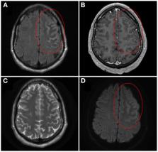
          Myelin oligodendrocyte glycoprotein antibody-associated disease (MOGAD) is an inflammatory demyelinating disease that is distinct from multiple sclerosis. Initial manifestations of MOGAD that were reported in the literature included optic neuritis, myelitis, brainstem demyelination and encephalitis, with emphasis placed on acute disseminated encephalomyelitis (ADEM) as the primary encephalitic presentation. In 2017, however, Ogawa et al. described four patients with seizures, unilateral cortical hyperintensities on brain magnetic resonance imaging T2-fluid-attenuated inversion recovery sequences, and anti-MOG positivity, indicating a potentially novel form of encephalitis in MOGAD. In 2019, we systematically reviewed the literature to better characterize this unique syndrome, which we referred to as unilateral cortical FLAIR-hyperintense Lesions in Anti-MOG-associated Encephalitis with Seizures (FLAMES). Subsequently, anti-MOG positivity in patients with a variety of cortical and meningeal disease presentations has been reported, indicating a broader spectrum of meningo-cortical manifestations in MOGAD that we review herein.

          Related collections

          Most cited references61

          • Record: found
          • Abstract: found
          • Article: not found

          A clinical approach to diagnosis of autoimmune encephalitis.

          Encephalitis is a severe inflammatory disorder of the brain with many possible causes and a complex differential diagnosis. Advances in autoimmune encephalitis research in the past 10 years have led to the identification of new syndromes and biomarkers that have transformed the diagnostic approach to these disorders. However, existing criteria for autoimmune encephalitis are too reliant on antibody testing and response to immunotherapy, which might delay the diagnosis. We reviewed the literature and gathered the experience of a team of experts with the aims of developing a practical, syndrome-based diagnostic approach to autoimmune encephalitis and providing guidelines to navigate through the differential diagnosis. Because autoantibody test results and response to therapy are not available at disease onset, we based the initial diagnostic approach on neurological assessment and conventional tests that are accessible to most clinicians. Through logical differential diagnosis, levels of evidence for autoimmune encephalitis (possible, probable, or definite) are achieved, which can lead to prompt immunotherapy.
            Bookmark
            • Record: found
            • Abstract: found
            • Article: not found

            Treatment and prognostic factors for long-term outcome in patients with anti-NMDA receptor encephalitis: an observational cohort study.

            Anti-NMDA receptor (NMDAR) encephalitis is an autoimmune disorder in which the use of immunotherapy and the long-term outcome have not been defined. We aimed to assess the presentation of the disease, the spectrum of symptoms, immunotherapies used, timing of improvement, and long-term outcome. In this multi-institutional observational study, we tested for the presence of NMDAR antibodies in serum or CSF samples of patients with encephalitis between Jan 1, 2007, and Jan 1, 2012. All patients who tested positive for NMDAR antibodies were included in the study; patients were assessed at symptom onset and at months 4, 8, 12, 18, and 24, by use of the modified Rankin scale (mRS). Treatment included first-line immunotherapy (steroids, intravenous immunoglobulin, plasmapheresis), second-line immunotherapy (rituximab, cyclophosphamide), and tumour removal. Predictors of outcome were determined at the Universities of Pennsylvania (PA, USA) and Barcelona (Spain) by use of a generalised linear mixed model with binary distribution. We enrolled 577 patients (median age 21 years, range 8 months to 85 years), 211 of whom were children (<18 years). Treatment effects and outcome were assessable in 501 (median follow-up 24 months, range 4-186): 472 (94%) underwent first-line immunotherapy or tumour removal, resulting in improvement within 4 weeks in 251 (53%). Of 221 patients who did not improve with first-line treatment, 125 (57%) received second-line immunotherapy that resulted in a better outcome (mRS 0-2) than those who did not (odds ratio [OR] 2·69, CI 1·24-5·80; p=0·012). During the first 24 months, 394 of 501 patients achieved a good outcome (mRS 0-2; median 6 months, IQR 2-12) and 30 died. At 24 months' follow-up, 203 (81%) of 252 patients had good outcome. Outcomes continued to improve for up to 18 months after symptom onset. Predictors of good outcome were early treatment (0·62, 0·50-0·76; p<0·0001) and no admission to an intensive care unit (0·12, 0·06-0·22; p<0·0001). 45 patients had one or multiple relapses (representing a 12% risk within 2 years); 46 (67%) of 69 relapses were less severe than initial episodes (p<0·0001). In 177 children, predictors of good outcome and the magnitude of effect of second-line immunotherapy were similar to those of the entire cohort. Most patients with anti-NMDAR encephalitis respond to immunotherapy. Second-line immunotherapy is usually effective when first-line treatments fail. In this cohort, the recovery of some patients took up to 18 months. The Dutch Cancer Society, the National Institutes of Health, the McKnight Neuroscience of Brain Disorders award, The Fondo de Investigaciones Sanitarias, and Fundació la Marató de TV3. Copyright © 2013 Elsevier Ltd. All rights reserved.
              Bookmark
              • Record: found
              • Abstract: found
              • Article: not found

              Antibody titres at diagnosis and during follow-up of anti-NMDA receptor encephalitis: a retrospective study.

              Anti-N-methyl-d-aspartate (NMDA) receptor encephalitis is a severe but treatable autoimmune disorder which diagnosis depends on sensitive and specific antibody testing. We aimed to assess the sensitivity and specificity of serum and CSF antibody testing in patients with anti-NMDA receptor encephalitis, and the relation between titres, relapses, outcome, and epitope repertoire. In this observational study, we used rat brain immunohistochemistry and cell-based assays (CBA) with fixed or live NMDA receptor-expressing cells to determine the sensitivity and specificity of antibody testing in paired serum and CSF samples. Samples were obtained at diagnosis from patients with anti-NMDA receptor encephalitis and from control participants worldwide. We deemed a patient to be antibody positive if their serum, their CSF, or both tested positive with both immunohistochemistry and CBA techniques; we determined titres with serial sample dilution using brain immunohistochemistry. We examined samples from 45 patients (25 with good outcome [modified Rankin Scale, mRS 0-2], ten with poor outcome [mRS 3-6], and ten with relapses) at three or more timepoints. We determined the epitope repertoire in the samples of 23 patients with CBA expressing GluN1-NMDA receptor mutants. We analysed samples from 250 patients with anti-NMDA receptor encephalitis and 100 control participants. All 250 patients had NMDA receptor antibodies in CSF but only 214 had antibodies in serum (sensitivity 100.0% [98.5-1000%] vs 85.6% [80.7-89.4%], p<0.0001). Serum immunohistochemistry testing was more often in agreement with CBA with fixed cells (77 [71%] of 108) than with CBA with live cells (63 [58%] of 108, p=0.0056). In multivariable analysis, CSF and serum titres were higher in patients with poor outcome than in those with good outcome (CSF dilution 340 vs 129, difference 211, [95% CI 1-421], p=0.049; serum dilution 7370 vs 1243, difference 6127 [2369-9885], p=0.0025), and in patients with teratoma than in those without teratoma (CSF 395 vs 110, difference 285 [134-437], p=0.0079; serum 5515 vs 1644, difference 3870 [548-7193], p=0.024). Over time there was a decrease of antibody titres in the 35 patients with good or poor outcome and samples followed at three timepoints regardless of outcome (from diagnosis to last follow-up: CSF 614 to 76, difference 538 [288-788]; serum 5460 to 1564, difference 3896 [2428-5362]; both p<0.0001). Relapses were associated with a change in titre more often in CSF than in serum (14 of 19 vs seven of 16, p=0.037). After recovery, 24 of 28 CSF samples and 17 of 23 serum samples from patients remained antibody positive. Patients' antibodies targeted a main epitope region at GluN1 aminoacid 369; the epitope repertoire did not differ between patients with different outcomes, and did not change during relapses. The sensitivity of NMDA receptor antibody testing is higher in CSF than in serum. Antibody titres in CSF and serum were higher in patients with poor outcome or teratoma than in patients with good outcome or no tumour. The titre change in CSF was more closely related with relapses than was that in serum. These findings emphasise the importance of including CSF in antibody studies, and that antibody titres can complement clinical assessments. Dutch Cancer Society, National Institutes of Health, McKnight Neuroscience of Brain Disorders award, the Fondo de Investigaciones Sanitarias, ErasmusMC fellowship, and Fundació la Marató de TV3. Copyright © 2014 Elsevier Ltd. All rights reserved.
                Bookmark

                Author and article information

                Contributors
                Journal
                Front Neurol
                Front Neurol
                Front. Neurol.
                Frontiers in Neurology
                Frontiers Media S.A.
                1664-2295
                20 October 2022
                2022
                : 13
                : 1044642
                Affiliations
                [1] 1Department of Clinical Neurological Sciences, Western University, London Health Sciences Centre , London, ON, Canada
                [2] 2Department of Pathology and Laboratory Medicine, Western University, London Health Sciences Centre , London, ON, Canada
                [3] 3Department of Medical Imaging, Western University, London Health Sciences Centre , London, ON, Canada
                Author notes

                Edited by: Sergio Muñiz-Castrillo, Stanford Center for Sleep Sciences and Medicine, United States

                Reviewed by: Giulia Fadda, McGill University, Canada; Mayra Montalvo Perero, University of Florida, United States

                *Correspondence: Adrian Budhram Adrian.budhram@ 123456lhsc.on.ca

                This article was submitted to Multiple Sclerosis and Neuroimmunology, a section of the journal Frontiers in Neurology

                Article
                10.3389/fneur.2022.1044642
                9630470
                36341089
                56bb32d1-63ef-42e7-87b3-36d2bd61cdfc
                Copyright © 2022 Budhram, Mirian and Sharma.

                This is an open-access article distributed under the terms of the Creative Commons Attribution License (CC BY). The use, distribution or reproduction in other forums is permitted, provided the original author(s) and the copyright owner(s) are credited and that the original publication in this journal is cited, in accordance with accepted academic practice. No use, distribution or reproduction is permitted which does not comply with these terms.

                History
                : 14 September 2022
                : 29 September 2022
                Page count
                Figures: 1, Tables: 1, Equations: 0, References: 61, Pages: 8, Words: 6012
                Categories
                Neurology
                Mini Review

                Neurology
                mog,flames,cerebral cortical encephalitis,meningitis,autoimmune neurology,neuroimmunology,mogad
                Neurology
                mog, flames, cerebral cortical encephalitis, meningitis, autoimmune neurology, neuroimmunology, mogad

                Comments

                Comment on this article