- Record: found
- Abstract: found
- Article: found
18F-FDG PET/CT with unusual bone and CNS metastases from testicular seminoma
research-article
Read this article at
There is no author summary for this article yet. Authors can add summaries to their articles on ScienceOpen to make them more accessible to a non-specialist audience.
Abstract
A 31 year old male with a previous history of testicular seminoma with complete reponse
after orchiectomy and three cycles of BEP scheme, was referred for 18F-FDG PET/CT
with a standard procedure for progressive decline consistent in spinal pain, gait
difficulty and Charcot’s neurologic triad (scanning speech, intention tremor and nystagmus)
initiated eight month after third course of chemotherapy. Dorsal spine MRI revealed
a space-occupying lesion at left T6 lamina. Histology examination confirmed a seminoma
metastatic to spine.
A wholebody and cerebral 18F-FDG PET/CT scan was performed 60 minutes after intravenous
injection of 370 MBq of 18F-FDG. PET/CT scan demonstrated an augmentation of soft
tissue due laminectomy with increased uptake of radiotracer and a Standardized Uptake
Value (SUV) maximum of 4.84 (Figure-1, Panel a), so persistence of tumour tissue cannot
be excluded. Furthermore, two focal hypermetabolic areas in CNS were revealed. First,
located in the spinal cord at C4-C5 vertebral levels with a SUV maximum of 7.49 (Figure-1,
Panel b) and second, in the cerebellum with a SUV maximum of 11 (Figure-1, Panel c),
corresponding with 3.9 cm mass in vermix observed at post-hoc MRI scan.
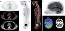
Figure 1
Wholebody and cerebral 18F-FDG PET/CT scan after i.v. administration of 370 MBq of
18F-FDG. Panel a) Axial images that revealed an augmentation of soft tissue due left
T6 laminectomy with increased uptake of radiotracer and SUV maximum of 4.84 that cannot
allow to excluded the persistence of tumour tissue. Panel b) Sagital images showed
a spinal cord metastasis with increased uptake of radiotracer at C4-C5 vertebral levels
with a SUV maximum of 7.49. Panel c) Cerebral scan revealed a hypermetabolic mass
in cerebellum, with high uptake of 18F-FDG (SUV maximum of 11), corresponding with
3.9 cm mass in vermix observed at post-hoc MRI scan.
This is an unusual intra-extracranial metastatic tumor merits active treatment.
Most relapses of seminoma occur within the first 3 years after orchiectomy. Bone and
CNS metastases involvement are an extremely rare event. A report of 2,550 patients
revealed bone metastases only in 3 patients with seminoma (0.12%) (1). Moreover, CNS
occurred only once in a series of 142 patients (0.7%) (2). Higher uptake in seminomas
than in nonseminomas testicular carcinomas (3) makes 18F-FDG PET/CT a powerful tool
in evaluating postchemotherapy seminoma relapses.
Related collections
Most cited references6
- Record: found
- Abstract: found
- Article: not found
Bone metastases in germ cell tumours: lessons learnt from a large retrospective study.
Mariam Jamal-Hanjani, Anna Karpathakis, Amy Kwan … (2013)
- Record: found
- Abstract: found
- Article: not found
FDG PET for detection and therapy control of metastatic germ cell tumor.
- Record: found
- Abstract: found
- Article: not found
Advanced seminoma: treatment results, survival, and prognostic factors in 142 patients.
P J Mencel, R Motzer, M. Mazumdar … (1994)
Author and article information
Comments
Comment on this article
scite_