25
views
0
recommends
+1 Recommend
0 collections
    0
    shares
      • Record: found
      • Abstract: found
      • Article: found
      Is Open Access

      Autophagy and the Cell Cycle: A Complex Landscape

      review-article

      Read this article at

      Bookmark
          There is no author summary for this article yet. Authors can add summaries to their articles on ScienceOpen to make them more accessible to a non-specialist audience.

          Abstract

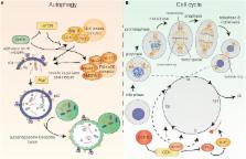
          Autophagy is a self-degradation pathway, in which cytoplasmic material is sequestered in double-membrane vesicles and delivered to the lysosome for degradation. Under basal conditions, autophagy plays a homeostatic function. However, in response to various stresses, the pathway can be further induced to mediate cytoprotection. Defective autophagy has been linked to a number of human pathologies, including neoplastic transformation, even though autophagy can also sustain the growth of tumor cells in certain contexts. In recent years, a considerable correlation has emerged between autophagy induction and stress-related cell-cycle responses, as well as unexpected roles for autophagy factors and selective autophagic degradation in the process of cell division. These advances have obvious implications for our understanding of the intricate relationship between autophagy and cancer. In this review, we will discuss our current knowledge of the reciprocal regulation connecting the autophagy pathway and cell-cycle progression. Furthermore, key findings involving nonautophagic functions for autophagy-related factors in cell-cycle regulation will be addressed.

          Related collections

          Most cited references114

          • Record: found
          • Abstract: found
          • Article: not found

          Guidelines for the use and interpretation of assays for monitoring autophagy.

          In 2008 we published the first set of guidelines for standardizing research in autophagy. Since then, research on this topic has continued to accelerate, and many new scientists have entered the field. Our knowledge base and relevant new technologies have also been expanding. Accordingly, it is important to update these guidelines for monitoring autophagy in different organisms. Various reviews have described the range of assays that have been used for this purpose. Nevertheless, there continues to be confusion regarding acceptable methods to measure autophagy, especially in multicellular eukaryotes. A key point that needs to be emphasized is that there is a difference between measurements that monitor the numbers or volume of autophagic elements (e.g., autophagosomes or autolysosomes) at any stage of the autophagic process vs. those that measure flux through the autophagy pathway (i.e., the complete process); thus, a block in macroautophagy that results in autophagosome accumulation needs to be differentiated from stimuli that result in increased autophagic activity, defined as increased autophagy induction coupled with increased delivery to, and degradation within, lysosomes (in most higher eukaryotes and some protists such as Dictyostelium) or the vacuole (in plants and fungi). In other words, it is especially important that investigators new to the field understand that the appearance of more autophagosomes does not necessarily equate with more autophagy. In fact, in many cases, autophagosomes accumulate because of a block in trafficking to lysosomes without a concomitant change in autophagosome biogenesis, whereas an increase in autolysosomes may reflect a reduction in degradative activity. Here, we present a set of guidelines for the selection and interpretation of methods for use by investigators who aim to examine macroautophagy and related processes, as well as for reviewers who need to provide realistic and reasonable critiques of papers that are focused on these processes. These guidelines are not meant to be a formulaic set of rules, because the appropriate assays depend in part on the question being asked and the system being used. In addition, we emphasize that no individual assay is guaranteed to be the most appropriate one in every situation, and we strongly recommend the use of multiple assays to monitor autophagy. In these guidelines, we consider these various methods of assessing autophagy and what information can, or cannot, be obtained from them. Finally, by discussing the merits and limits of particular autophagy assays, we hope to encourage technical innovation in the field.
            Bookmark
            • Record: found
            • Abstract: found
            • Article: not found

            Non-muscle myosin II takes centre stage in cell adhesion and migration.

            Non-muscle myosin II (NM II) is an actin-binding protein that has actin cross-linking and contractile properties and is regulated by the phosphorylation of its light and heavy chains. The three mammalian NM II isoforms have both overlapping and unique properties. Owing to its position downstream of convergent signalling pathways, NM II is central in the control of cell adhesion, cell migration and tissue architecture. Recent insight into the role of NM II in these processes has been gained from loss-of-function and mutant approaches, methods that quantitatively measure actin and adhesion dynamics and the discovery of NM II mutations that cause monogenic diseases.
              Bookmark
              • Record: found
              • Abstract: not found
              • Article: not found

              CDK inhibitors: positive and negative regulators of G1-phase progression.

                Bookmark

                Author and article information

                Contributors
                URI : http://frontiersin.org/people/u/414536
                URI : http://frontiersin.org/people/u/411332
                URI : http://frontiersin.org/people/u/423898
                Journal
                Front Oncol
                Front Oncol
                Front. Oncol.
                Frontiers in Oncology
                Frontiers Media S.A.
                2234-943X
                31 March 2017
                2017
                : 7
                : 51
                Affiliations
                [1] 1Cell Stress and Survival Unit, Danish Cancer Society Research Center , Copenhagen, Denmark
                [2] 2Department of Biology, University of Rome Tor Vergata , Rome, Italy
                [3] 3Department of Pediatric Hematology and Oncology, Istituto di Ricovero e Cura a Carattere Scientifico Bambino Gesù Children’s Hospital , Rome, Italy
                Author notes

                Edited by: Jon Lane, University of Bristol, UK

                Reviewed by: Viktor Korolchuk, Newcastle University, UK; Simon Wilkinson, University of Edinburgh, UK

                *Correspondence: Daniela De Zio, dzio@ 123456cancer.dk ; Francesco Cecconi, cecconi@ 123456cancer.dk

                Specialty section: This article was submitted to Molecular and Cellular Oncology, a section of the journal Frontiers in Oncology

                Article
                10.3389/fonc.2017.00051
                5374984
                28409123
                54dead82-5499-4958-9997-0196ee9f201e
                Copyright © 2017 Mathiassen, De Zio and Cecconi.

                This is an open-access article distributed under the terms of the Creative Commons Attribution License (CC BY). The use, distribution or reproduction in other forums is permitted, provided the original author(s) or licensor are credited and that the original publication in this journal is cited, in accordance with accepted academic practice. No use, distribution or reproduction is permitted which does not comply with these terms.

                History
                : 01 February 2017
                : 10 March 2017
                Page count
                Figures: 2, Tables: 0, Equations: 0, References: 171, Pages: 16, Words: 13947
                Funding
                Funded by: Fondazione Telethon 10.13039/501100002426
                Award ID: GGP14202
                Funded by: Associazione Italiana per la Ricerca sul Cancro 10.13039/501100005010
                Award ID: IG2016-18906
                Funded by: Fondazione Italiana Sclerosi Multipla 10.13039/100007366
                Award ID: 2013
                Funded by: Kræftens Bekæmpelse 10.13039/100008363
                Award ID: R146-A9364
                Funded by: Novo Nordisk 10.13039/501100004191
                Award ID: 7559, 22544
                Funded by: Horizon 2020 Framework Programme 10.13039/100010661
                Award ID: 642295
                Funded by: Danmarks Grundforskningsfond 10.13039/501100001732
                Award ID: CARD
                Categories
                Oncology
                Review

                Oncology & Radiotherapy
                autophagy,cancer,cell cycle,cell stress,senescence,mitosis,cytokinesis,p53
                Oncology & Radiotherapy
                autophagy, cancer, cell cycle, cell stress, senescence, mitosis, cytokinesis, p53

                Comments

                Comment on this article