1
views
0
recommends
+1 Recommend
0 collections
    0
    shares
      • Record: found
      • Abstract: found
      • Article: found
      Is Open Access

      Role of echography in diagnostic dilemma in choroidal masses

      research-article

      Read this article at

      Bookmark
          There is no author summary for this article yet. Authors can add summaries to their articles on ScienceOpen to make them more accessible to a non-specialist audience.

          Abstract

          Purpose:

          To evaluate the role of echography in diagnosis and management of a diverse array of choroidal masses.

          Materials and Methods:

          Sixty-two cases of clinically suspected choroidal masses were prospectively analyzed with B-scan (10 Hz), A-scan, and ultrasound biomicroscopy (UBM) (50 Hz) after a meticulous history and ocular examination. Ancillary investigations and systemic evaluation were also done.

          Results:

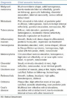
          Based on clinical suspicion, acoustic features, response to treatment, and other ancillary tests combined together, the various masses were differentiated. The cases included in the study were as follows: n = 10 malignant melanomas, n = 16 metastasis and infiltrations, n = 9 hemangioma, n = 7 tuberculoma, n = 8 nonspecific inflammatory masses, n = 2 disciform plaques, n = 4 macular cysts or retinoschisis, n = 2 Coat's disease, n = 1 melanocytoma, and n = 2 osteomas. Ultrasonography (USG) alone could identify n = 51 lesions, while UBM in combination with USG was needed in remaining 11 masses.

          Conclusion:

          Standardized echography is an important adjunct in the diagnosis and management of eyes with intraocular masses. A better understanding of the clinicopathological and echographic picture of the diverse lesions can help in detection, differentiation, diagnosis, proposing a therapeutic approach, and also monitoring response to treatment. Echography is essential to evaluate tumors for extrascleral and anterior segment extension.

          Related collections

          Most cited references10

          • Record: found
          • Abstract: found
          • Article: not found

          Pseudomelanomas of the posterior uveal tract: the 2006 Taylor R. Smith Lecture.

          To determine the types and frequency of lesions that clinically simulate choroidal or ciliary body melanoma (posterior uveal melanoma; PUM). A review was conducted on cases of patients referred to the ocular oncology service from October 1978 through September 2003 with the diagnosis of possible PUM but who were subsequently diagnosed by the authors to have a simulating lesion rather than PUM. The type and percent of pseudomelanomas were tabulated and compared with findings of a similar study from our service on data collected before 1978. There were approximately 12,000 patients referred because of a lesion believed to be a PUM during the 25 years included in the data collection. Of these patients, 1,739 (14%) were found to have a simulating condition. There were 54 different conditions that simulated melanoma. The most frequent condition was choroidal nevus, accounting for 851 cases (49%) of the pseudomelanomas. This was followed by peripheral exudative hemorrhagic chorioretinopathy (139 cases; 8%), congenital hypertrophy of the retinal pigment epithelium (108 cases; 6%), hemorrhagic detachment of the retina or pigment epithelium (86 cases; 5%), circumscribed choroidal hemangioma (79 cases; 5%) and age-related macular degeneration (76 cases; 4%). Compared with the 1980 report, the rate of pseudomelanomas diagnosed as choroidal nevus increased from 26% to 49%. A variety of lesions can simulate PUM. Suspicious choroidal nevus is still the lesion most difficult to differentiate from PUM. Most other pseudomelanomas account for a lower percent compared with findings from the prior study, suggesting that clinicians are now more familiar with the other pseudomelanomas and less likely to refer them to rule out PUM.
            Bookmark
            • Record: found
            • Abstract: found
            • Article: found
            Is Open Access

            Pictorial essay: B-scan ultrasonography in ocular abnormalities

            B-scan ultrasonography (USG) is a simple, noninvasive tool for diagnosing lesions of the posterior segment of the eyeball. Common conditions such as cataract, vitreous degeneration, retinal detachment, ocular trauma, choroidal melanoma, and retinoblastoma can be accurately evaluated with this modality. B-scan USG is cost-effective, which is an important consideration in the rural setting. In addition, it is noninvasive and easily available and the results are reproducible.
              Bookmark
              • Record: found
              • Abstract: not found
              • Article: not found

              Ultrasonic diagnosis of tumors of the choroid.

                Bookmark

                Author and article information

                Journal
                Indian J Ophthalmol
                Indian J Ophthalmol
                IJO
                Indian Journal of Ophthalmology
                Medknow Publications & Media Pvt Ltd (India )
                0301-4738
                1998-3689
                February 2014
                : 62
                : 2
                : 167-170
                Affiliations
                [1]LVPEI, Hyderabad, India
                [1 ]Ultrasound and Retina Department, M&J Institute, Ophthalmology, Ahmedabad, Gujarat, India
                Author notes
                Correspondence to: Prof. Hansa H. Thakkar, Ultrasound and Retina Department, M&J Institute of Ophthalmology, Ahmedabad, Gujarat, India. E-mail: dr.hansathakkar@ 123456gmail.com
                Article
                IJO-62-167
                10.4103/0301-4738.128626
                4005232
                24618487
                54712273-40a8-4db4-ad25-1c56d4c0ff52
                Copyright: © Indian Journal of Ophthalmology

                This is an open-access article distributed under the terms of the Creative Commons Attribution-Noncommercial-Share Alike 3.0 Unported, which permits unrestricted use, distribution, and reproduction in any medium, provided the original work is properly cited.

                History
                : 11 August 2012
                : 16 September 2013
                Categories
                Original Article

                Ophthalmology & Optometry
                acoustic features,a-scan,b-scan,choroidal mass,ultrasound biomicroscopy
                Ophthalmology & Optometry
                acoustic features, a-scan, b-scan, choroidal mass, ultrasound biomicroscopy

                Comments

                Comment on this article