2
views
0
recommends
+1 Recommend
0 collections
    0
    shares
      • Record: found
      • Abstract: found
      • Article: found
      Is Open Access

      Cisplatin-Induced Stria Vascularis Damage Is Associated with Inflammation and Fibrosis

      research-article
      , , , ,
      Neural Plasticity
      Hindawi

      Read this article at

      Bookmark
          There is no author summary for this article yet. Authors can add summaries to their articles on ScienceOpen to make them more accessible to a non-specialist audience.

          Abstract

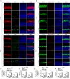
          The stria vascularis (SV) generates the endocochlear potential (EP) in the inner ear and is necessary for proper hair cell (HC) mechanotransduction and hearing. Cell junctions are indispensable for the establishment of compositionally distinct fluid compartments in the inner ear. Ototoxic drug cisplatin can damage SV and cause sensorineural hearing loss; however, the underlying mechanisms behind such injury are unclear. In this study, after the intraperitoneal injection of cisplatin (3 mg/kg/day for 7 days) in mice, we determined the auditory function by EP recording and auditory brainstem response (ABR) analysis, observed the ultrastructure of SV by transmission electron microscopy (TEM), and examined the expression and distribution of cell junction proteins by western blot, PCR, and immunofluorescence staining. We discovered that the EP was significantly reduced while ABR thresholds were significantly elevated in cisplatin-treated mice; cisplatin induced ultrastructural changes in marginal cells (MCs), endothelial cells (ECs), pericytes, etc. We found that cisplatin insulted auditory function not only by reducing the expression of zonula occludens protein-1 (ZO-1) in MCs of the SV but also by decreasing the expression of connexin 26 (Cx26) and connexin 43 (Cx43) in MCs and basal cells (BCs). More importantly, cisplatin induced activations of perivascular-resident macrophage-like melanocytes (PVM/Ms) and interleukin-1beta (IL-1 β) as well as increased expressions of profibrotic proteins such as laminin and collagen IV in SV. Thus, our results firstly showed that cisplatin induced fibrosis, inflammation, and the complex expression change of cell junctions in SV.

          Related collections

          Most cited references56

          • Record: found
          • Abstract: found
          • Article: not found

          Macrophage-to-Myofibroblast Transition Contributes to Interstitial Fibrosis in Chronic Renal Allograft Injury.

          Interstitial fibrosis is an important contributor to graft loss in chronic renal allograft injury. Inflammatory macrophages are associated with fibrosis in renal allografts, but how these cells contribute to this damaging response is not clearly understood. Here, we investigated the role of macrophage-to-myofibroblast transition in interstitial fibrosis in human and experimental chronic renal allograft injury. In biopsy specimens from patients with active chronic allograft rejection, we identified cells undergoing macrophage-to-myofibroblast transition by the coexpression of macrophage (CD68) and myofibroblast (α-smooth muscle actin [α-SMA]) markers. CD68(+)/α-SMA(+) cells accounted for approximately 50% of the myofibroblast population, and the number of these cells correlated with allograft function and the severity of interstitial fibrosis. Similarly, in C57BL/6J mice with a BALB/c renal allograft, cells coexpressing macrophage markers (CD68 or F4/80) and α-SMA composed a significant population in the interstitium of allografts undergoing chronic rejection. Fate-mapping in Lyz2-Cre/Rosa26-Tomato mice showed that approximately half of α-SMA(+) myofibroblasts in renal allografts originated from recipient bone marrow-derived macrophages. Knockout of Smad3 protected against interstitial fibrosis in renal allografts and substantially reduced the number of macrophage-to-myofibroblast transition cells. Furthermore, the majority of macrophage-to-myofibroblast transition cells in human and experimental renal allograft rejection coexpressed the M2-type macrophage marker CD206, and this expression was considerably reduced in Smad3-knockout recipients. In conclusion, our studies indicate that macrophage-to-myofibroblast transition contributes to interstitial fibrosis in chronic renal allograft injury. Moreover, the transition of bone marrow-derived M2-type macrophages to myofibroblasts in the renal allograft is regulated via a Smad3-dependent mechanism.
            Bookmark
            • Record: found
            • Abstract: found
            • Article: found
            Is Open Access

            Autophagy protects auditory hair cells against neomycin-induced damage

            ABSTRACT Aminoglycosides are toxic to sensory hair cells (HCs). Macroautophagy/autophagy is an essential and highly conserved self-digestion pathway that plays important roles in the maintenance of cellular function and viability under stress. However, the role of autophagy in aminoglycoside-induced HC injury is unknown. Here, we first found that autophagy activity was significantly increased, including enhanced autophagosome-lysosome fusion, in both cochlear HCs and HEI-OC-1 cells after neomycin or gentamicin injury, suggesting that autophagy might be correlated with aminoglycoside-induced cell death. We then used rapamycin, an autophagy activator, to increase the autophagy activity and found that the ROS levels, apoptosis, and cell death were significantly decreased after neomycin or gentamicin injury. In contrast, treatment with the autophagy inhibitor 3-methyladenine (3-MA) or knockdown of autophagy-related (ATG) proteins resulted in reduced autophagy activity and significantly increased ROS levels, apoptosis, and cell death after neomycin or gentamicin injury. Finally, after neomycin injury, the antioxidant N-acetylcysteine could successfully prevent the increased apoptosis and HC loss induced by 3-MA treatment or ATG knockdown, suggesting that autophagy protects against neomycin-induced HC damage by inhibiting oxidative stress. We also found that the dysfunctional mitochondria were not eliminated by selective autophagy (mitophagy) in HEI-OC-1 cells after neomycin treatment, suggesting that autophagy might not directly target the damaged mitochondria for degradation. This study demonstrates that moderate ROS levels can promote autophagy to recycle damaged cellular constituents and maintain cellular homeostasis, while the induction of autophagy can inhibit apoptosis and protect the HCs by suppressing ROS accumulation after aminoglycoside injury.
              Bookmark
              • Record: found
              • Abstract: found
              • Article: not found

              Spontaneous hair cell regeneration in the neonatal mouse cochlea in vivo.

              Loss of cochlear hair cells in mammals is currently believed to be permanent, resulting in hearing impairment that affects more than 10% of the population. Here, we developed two genetic strategies to ablate neonatal mouse cochlear hair cells in vivo. Both Pou4f3(DTR/+) and Atoh1-CreER™; ROSA26(DTA/+) alleles allowed selective and inducible hair cell ablation. After hair cell loss was induced at birth, we observed spontaneous regeneration of hair cells. Fate-mapping experiments demonstrated that neighboring supporting cells acquired a hair cell fate, which increased in a basal to apical gradient, averaging over 120 regenerated hair cells per cochlea. The normally mitotically quiescent supporting cells proliferated after hair cell ablation. Concurrent fate mapping and labeling with mitotic tracers showed that regenerated hair cells were derived by both mitotic regeneration and direct transdifferentiation. Over time, regenerated hair cells followed a similar pattern of maturation to normal hair cell development, including the expression of prestin, a terminal differentiation marker of outer hair cells, although many new hair cells eventually died. Hair cell regeneration did not occur when ablation was induced at one week of age. Our findings demonstrate that the neonatal mouse cochlea is capable of spontaneous hair cell regeneration after damage in vivo. Thus, future studies on the neonatal cochlea might shed light on the competence of supporting cells to regenerate hair cells and on the factors that promote the survival of newly regenerated hair cells.
                Bookmark

                Author and article information

                Contributors
                Journal
                Neural Plast
                Neural Plast
                NP
                Neural Plasticity
                Hindawi
                2090-5904
                1687-5443
                2020
                22 September 2020
                : 2020
                : 8851525
                Affiliations
                Department of Otolaryngology-Head and Neck Surgery, Shandong Provincial ENT Hospital, Cheeloo College of Medicine, Shandong University, Jinan 250022, China
                Author notes

                Academic Editor: Jian Wang

                Author information
                https://orcid.org/0000-0002-8509-6042
                https://orcid.org/0000-0003-0955-9532
                https://orcid.org/0000-0001-6031-7972
                Article
                10.1155/2020/8851525
                7527906
                33029120
                54287037-a8fc-4fb4-ac0d-7248b1c9cb12
                Copyright © 2020 Na Zhang et al.

                This is an open access article distributed under the Creative Commons Attribution License, which permits unrestricted use, distribution, and reproduction in any medium, provided the original work is properly cited.

                History
                : 19 April 2020
                : 5 August 2020
                : 3 September 2020
                Funding
                Funded by: Key Project of Shandong Provincial Programs for Research and Development
                Award ID: 2017CXGC1213
                Funded by: National Natural Science Foundation of China
                Award ID: 81670932
                Award ID: 81700918
                Funded by: Taishan Scholar Project of Shandong Province
                Award ID: ts20130913
                Award ID: tsqn201909189
                Categories
                Research Article

                Neurosciences
                Neurosciences

                Comments

                Comment on this article