6
views
0
recommends
+1 Recommend
0 collections
    0
    shares
      • Record: found
      • Abstract: found
      • Article: found
      Is Open Access

      Assessment of safety margin after microwave ablation of stage I NSCLC with three-dimensional reconstruction technique using CT imaging

      research-article

      Read this article at

      Bookmark
          There is no author summary for this article yet. Authors can add summaries to their articles on ScienceOpen to make them more accessible to a non-specialist audience.

          Abstract

          Objective

          To assess the ablative margin of microwave ablation (MWA) for stage I non-small cell lung cancer (NSCLC) using a three-dimensional (3D) reconstruction technique.

          Materials and methods

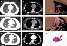
          We retrospectively analyzed 36 patients with stage I NSCLC lesions undergoing MWA and analyzed the relationship between minimal ablative margin and the local tumor progression (LTP) interval, the distant metastasis interval and disease-free survival (DFS). The minimal ablative margin was measured using the fusion of 3D computed tomography reconstruction technique.

          Results

          Univariate and multivariate analyses indicated that tumor size (hazard ratio [HR] = 1.91, P < 0.01; HR = 2.41, P = 0.01) and minimal ablative margin (HR = 0.13, P < 0.01; HR = 0.11, P < 0.01) were independent prognostic factors for the LTP interval. Tumor size (HR = 1.96, P < 0.01; HR = 2.35, P < 0.01) and minimal ablative margin (HR = 0.17, P < 0.01; HR = 0.13, P < 0.01) were independent prognostic factors for DFS by univariate and multivariate analyses. In the group with a minimal ablative margin < 5 mm, the 1-year and 2-year local progression-free rates were 35.7% and 15.9%, respectively. The 1-year and 2-year distant metastasis-free rates were 75.6% and 75.6%, respectively; the 1-year and 2-year disease-free survival rates were 16.7% and 11.1%, respectively. In the group with a minimal ablative margin ≥ 5 mm, the 1-year and 2-year local progression-free rates were 88.9% and 69.4%, respectively. The 1-year and 2-year distant metastasis-free rates were 94.4% and 86.6%, respectively; the 1-year and 2-year disease-free survival rates were 88.9% and 63.7%, respectively. The feasibility of 3D quantitative analysis of the ablative margins after MWA for NSCLC has been validated.

          Conclusions

          The minimal ablative margin is an independent factor of NSCLC relapse after MWA, and the fusion of 3D reconstruction technique can feasibly assess the minimal ablative margin.

          Supplementary Information

          The online version contains supplementary material available at 10.1186/s12880-021-00626-z.

          Related collections

          Most cited references32

          • Record: found
          • Abstract: found
          • Article: not found

          Global Cancer Statistics 2018: GLOBOCAN Estimates of Incidence and Mortality Worldwide for 36 Cancers in 185 Countries

          This article provides a status report on the global burden of cancer worldwide using the GLOBOCAN 2018 estimates of cancer incidence and mortality produced by the International Agency for Research on Cancer, with a focus on geographic variability across 20 world regions. There will be an estimated 18.1 million new cancer cases (17.0 million excluding nonmelanoma skin cancer) and 9.6 million cancer deaths (9.5 million excluding nonmelanoma skin cancer) in 2018. In both sexes combined, lung cancer is the most commonly diagnosed cancer (11.6% of the total cases) and the leading cause of cancer death (18.4% of the total cancer deaths), closely followed by female breast cancer (11.6%), prostate cancer (7.1%), and colorectal cancer (6.1%) for incidence and colorectal cancer (9.2%), stomach cancer (8.2%), and liver cancer (8.2%) for mortality. Lung cancer is the most frequent cancer and the leading cause of cancer death among males, followed by prostate and colorectal cancer (for incidence) and liver and stomach cancer (for mortality). Among females, breast cancer is the most commonly diagnosed cancer and the leading cause of cancer death, followed by colorectal and lung cancer (for incidence), and vice versa (for mortality); cervical cancer ranks fourth for both incidence and mortality. The most frequently diagnosed cancer and the leading cause of cancer death, however, substantially vary across countries and within each country depending on the degree of economic development and associated social and life style factors. It is noteworthy that high-quality cancer registry data, the basis for planning and implementing evidence-based cancer control programs, are not available in most low- and middle-income countries. The Global Initiative for Cancer Registry Development is an international partnership that supports better estimation, as well as the collection and use of local data, to prioritize and evaluate national cancer control efforts. CA: A Cancer Journal for Clinicians 2018;0:1-31. © 2018 American Cancer Society.
            Bookmark
            • Record: found
            • Abstract: found
            • Article: not found

            Cancer statistics, 2019

            Each year, the American Cancer Society estimates the numbers of new cancer cases and deaths that will occur in the United States and compiles the most recent data on cancer incidence, mortality, and survival. Incidence data, available through 2015, were collected by the Surveillance, Epidemiology, and End Results Program; the National Program of Cancer Registries; and the North American Association of Central Cancer Registries. Mortality data, available through 2016, were collected by the National Center for Health Statistics. In 2019, 1,762,450 new cancer cases and 606,880 cancer deaths are projected to occur in the United States. Over the past decade of data, the cancer incidence rate (2006-2015) was stable in women and declined by approximately 2% per year in men, whereas the cancer death rate (2007-2016) declined annually by 1.4% and 1.8%, respectively. The overall cancer death rate dropped continuously from 1991 to 2016 by a total of 27%, translating into approximately 2,629,200 fewer cancer deaths than would have been expected if death rates had remained at their peak. Although the racial gap in cancer mortality is slowly narrowing, socioeconomic inequalities are widening, with the most notable gaps for the most preventable cancers. For example, compared with the most affluent counties, mortality rates in the poorest counties were 2-fold higher for cervical cancer and 40% higher for male lung and liver cancers during 2012-2016. Some states are home to both the wealthiest and the poorest counties, suggesting the opportunity for more equitable dissemination of effective cancer prevention, early detection, and treatment strategies. A broader application of existing cancer control knowledge with an emphasis on disadvantaged groups would undoubtedly accelerate progress against cancer.
              Bookmark
              • Record: found
              • Abstract: found
              • Article: not found

              Image-guided tumor ablation: standardization of terminology and reporting criteria--a 10-year update.

              Image-guided tumor ablation has become a well-established hallmark of local cancer therapy. The breadth of options available in this growing field increases the need for standardization of terminology and reporting criteria to facilitate effective communication of ideas and appropriate comparison among treatments that use different technologies, such as chemical (eg, ethanol or acetic acid) ablation, thermal therapies (eg, radiofrequency, laser, microwave, focused ultrasound, and cryoablation) and newer ablative modalities such as irreversible electroporation. This updated consensus document provides a framework that will facilitate the clearest communication among investigators regarding ablative technologies. An appropriate vehicle is proposed for reporting the various aspects of image-guided ablation therapy including classification of therapies, procedure terms, descriptors of imaging guidance, and terminology for imaging and pathologic findings. Methods are addressed for standardizing reporting of technique, follow-up, complications, and clinical results. As noted in the original document from 2003, adherence to the recommendations will improve the precision of communications in this field, leading to more accurate comparison of technologies and results, and ultimately to improved patient outcomes. Online supplemental material is available for this article . © RSNA, 2014.
                Bookmark

                Author and article information

                Contributors
                154247850@qq.com
                Journal
                BMC Med Imaging
                BMC Med Imaging
                BMC Medical Imaging
                BioMed Central (London )
                1471-2342
                7 June 2021
                7 June 2021
                2021
                : 21
                : 96
                Affiliations
                [1 ]GRID grid.452222.1, Department of Oncology, , Jinan Central Hospital Affiliated to Shandong University, ; Jinan, People’s Republic of China
                [2 ]Department of Radiation, The 960th Hospital of the PLA Joint Logistics Support Force, Jinan, People’s Republic of China
                [3 ]GRID grid.452222.1, Department of Radiology, , Jinan Central Hospital Affiliated to Shandong University, ; Jinan, People’s Republic of China
                [4 ]GRID grid.412521.1, Department of Radiology, , The Affiliated Hospital of Qingdao University, ; Qingdao, People’s Republic of China
                Article
                626
                10.1186/s12880-021-00626-z
                8185913
                34098894
                53f329b5-2f90-42f4-abd3-51c18a97b891
                © The Author(s) 2021

                Open AccessThis article is licensed under a Creative Commons Attribution 4.0 International License, which permits use, sharing, adaptation, distribution and reproduction in any medium or format, as long as you give appropriate credit to the original author(s) and the source, provide a link to the Creative Commons licence, and indicate if changes were made. The images or other third party material in this article are included in the article's Creative Commons licence, unless indicated otherwise in a credit line to the material. If material is not included in the article's Creative Commons licence and your intended use is not permitted by statutory regulation or exceeds the permitted use, you will need to obtain permission directly from the copyright holder. To view a copy of this licence, visit http://creativecommons.org/licenses/by/4.0/. The Creative Commons Public Domain Dedication waiver ( http://creativecommons.org/publicdomain/zero/1.0/) applies to the data made available in this article, unless otherwise stated in a credit line to the data.

                History
                : 17 February 2021
                : 27 May 2021
                Categories
                Research
                Custom metadata
                © The Author(s) 2021

                Radiology & Imaging
                lung cancer,microwave ablation,ablative margin,three-dimensional reconstruction

                Comments

                Comment on this article