14
views
0
recommends
+1 Recommend
0 collections
    0
    shares
      • Record: found
      • Abstract: found
      • Article: found
      Is Open Access

      Crystal structure of SEL1L: Insight into the roles of SLR motifs in ERAD pathway

      research-article

      Read this article at
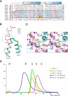
      Bookmark
          There is no author summary for this article yet. Authors can add summaries to their articles on ScienceOpen to make them more accessible to a non-specialist audience.

          Abstract

          Terminally misfolded proteins are selectively recognized and cleared by the endoplasmic reticulum-associated degradation (ERAD) pathway. SEL1L, a component of the ERAD machinery, plays an important role in selecting and transporting ERAD substrates for degradation. We have determined the crystal structure of the mouse SEL1L central domain comprising five Sel1- Like Repeats (SLR motifs 5 to 9; hereafter called SEL1L cent). Strikingly, SEL1L cent forms a homodimer with two-fold symmetry in a head-to-tail manner. Particularly, the SLR motif 9 plays an important role in dimer formation by adopting a domain-swapped structure and providing an extensive dimeric interface. We identified that the full-length SEL1L forms a self-oligomer through the SEL1L cent domain in mammalian cells. Furthermore, we discovered that the SLR-C, comprising SLR motifs 10 and 11, of SEL1L directly interacts with the N-terminus luminal loops of HRD1. Therefore, we propose that certain SLR motifs of SEL1L play a unique role in membrane bound ERAD machinery.

          Related collections

          Most cited references29

          • Record: found
          • Abstract: found
          • Article: not found

          Road to ruin: targeting proteins for degradation in the endoplasmic reticulum.

          Some nascent proteins that fold within the endoplasmic reticulum (ER) never reach their native state. Misfolded proteins are removed from the folding machinery, dislocated from the ER into the cytosol, and degraded in a series of pathways collectively referred to as ER-associated degradation (ERAD). Distinct ERAD pathways centered on different E3 ubiquitin ligases survey the range of potential substrates. We now know many of the components of the ERAD machinery and pathways used to detect substrates and target them for degradation. Much less is known about the features used to identify terminally misfolded conformations and the broader role of these pathways in regulating protein half-lives.
            Bookmark
            • Record: found
            • Abstract: found
            • Article: not found

            Defining human ERAD networks through an integrative mapping strategy

            SUMMARY Proteins that fail to correctly fold or assemble into oligomeric complexes in the endoplasmic reticulum (ER) are degraded by a ubiquitin and proteasome dependent process known as ER-associated degradation (ERAD). Although many individual components of the ERAD system have been identified, how these proteins are organised into a functional network that coordinates recognition, ubiquitination, and dislocation of substrates across the ER membrane is not well understood. We have investigated the functional organisation of the mammalian ERAD system using a systems-level strategy that integrates proteomics, functional genomics, and the transcriptional response to ER stress. This analysis supports an adaptive organisation for the mammalian ERAD machinery and reveals a number of metazoan-specific genes not previously linked to ERAD.
              Bookmark
              • Record: found
              • Abstract: found
              • Article: not found

              OS-9 and GRP94 deliver mutant alpha1-antitrypsin to the Hrd1-SEL1L ubiquitin ligase complex for ERAD.

              Terminally misfolded or unassembled proteins in the early secretory pathway are degraded by a ubiquitin- and proteasome-dependent process known as ER-associated degradation (ERAD). How substrates of this pathway are recognized within the ER and delivered to the cytoplasmic ubiquitin-conjugating machinery is unknown. We report here that OS-9 and XTP3-B/Erlectin are ER-resident glycoproteins that bind to ERAD substrates and, through the SEL1L adaptor, to the ER-membrane-embedded ubiquitin ligase Hrd1. Both proteins contain conserved mannose 6-phosphate receptor homology (MRH) domains, which are required for interaction with SEL1L, but not with substrate. OS-9 associates with the ER chaperone GRP94 which, together with Hrd1 and SEL1L, is required for the degradation of an ERAD substrate, mutant alpha(1)-antitrypsin. These data suggest that XTP3-B and OS-9 are components of distinct, partially redundant, quality control surveillance pathways that coordinate protein folding with membrane dislocation and ubiquitin conjugation in mammalian cells.
                Bookmark

                Author and article information

                Journal
                Sci Rep
                Sci Rep
                Scientific Reports
                Nature Publishing Group
                2045-2322
                09 February 2016
                2016
                : 6
                : 20261
                Affiliations
                [1 ]Department of Biological Sciences, School of Life Sciences, Ulsan National Institute of Science and Technology , 50 UNIST-gil, Ulsan 44919, Republic of Korea
                [2 ]Pohang Accelerator Laboratory, Pohang University of Science and Technology , Pohang, Kyungbuk 37673, Korea
                [3 ]School of Life Sciences, Gwangju Institute of Science and Technology , Gwangju 61005, Korea
                Author notes
                Article
                srep20261
                10.1038/srep20261
                4746701
                27064360
                534cc032-ca29-4bb9-b34f-ba670b7cb1a3
                Copyright © 2016, Macmillan Publishers Limited

                This work is licensed under a Creative Commons Attribution 4.0 International License. The images or other third party material in this article are included in the article’s Creative Commons license, unless indicated otherwise in the credit line; if the material is not included under the Creative Commons license, users will need to obtain permission from the license holder to reproduce the material. To view a copy of this license, visit http://creativecommons.org/licenses/by/4.0/

                History
                : 20 October 2015
                : 31 December 2015
                Categories
                Article

                Uncategorized
                Uncategorized

                Comments

                Comment on this article