169
views
0
recommends
+1 Recommend
1 collections
    0
    shares
      • Record: found
      • Abstract: found
      • Article: not found

      Radiological findings from 81 patients with COVID-19 pneumonia in Wuhan, China: a descriptive study

      research-article

      Read this article at

      ScienceOpenPublisherPMC
      Bookmark
          There is no author summary for this article yet. Authors can add summaries to their articles on ScienceOpen to make them more accessible to a non-specialist audience.

          Summary

          Background

          A cluster of patients with coronavirus disease 2019 (COVID-19) pneumonia caused by infection with severe acute respiratory syndrome coronavirus 2 (SARS-CoV-2) were successively reported in Wuhan, China. We aimed to describe the CT findings across different timepoints throughout the disease course.

          Methods

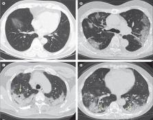
          Patients with COVID-19 pneumonia (confirmed by next-generation sequencing or RT-PCR) who were admitted to one of two hospitals in Wuhan and who underwent serial chest CT scans were retrospectively enrolled. Patients were grouped on the basis of the interval between symptom onset and the first CT scan: group 1 (subclinical patients; scans done before symptom onset), group 2 (scans done ≤1 week after symptom onset), group 3 (>1 week to 2 weeks), and group 4 (>2 weeks to 3 weeks). Imaging features and their distribution were analysed and compared across the four groups.

          Findings

          81 patients admitted to hospital between Dec 20, 2019, and Jan 23, 2020, were retrospectively enrolled. The cohort included 42 (52%) men and 39 (48%) women, and the mean age was 49·5 years (SD 11·0). The mean number of involved lung segments was 10·5 (SD 6·4) overall, 2·8 (3·3) in group 1, 11·1 (5·4) in group 2, 13·0 (5·7) in group 3, and 12·1 (5·9) in group 4. The predominant pattern of abnormality observed was bilateral (64 [79%] patients), peripheral (44 [54%]), ill-defined (66 [81%]), and ground-glass opacification (53 [65%]), mainly involving the right lower lobes (225 [27%] of 849 affected segments). In group 1 (n=15), the predominant pattern was unilateral (nine [60%]) and multifocal (eight [53%]) ground-glass opacities (14 [93%]). Lesions quickly evolved to bilateral (19 [90%]), diffuse (11 [52%]) ground-glass opacity predominance (17 [81%]) in group 2 (n=21). Thereafter, the prevalence of ground-glass opacities continued to decrease (17 [57%] of 30 patients in group 3, and five [33%] of 15 in group 4), and consolidation and mixed patterns became more frequent (12 [40%] in group 3, eight [53%] in group 4).

          Interpretation

          COVID-19 pneumonia manifests with chest CT imaging abnormalities, even in asymptomatic patients, with rapid evolution from focal unilateral to diffuse bilateral ground-glass opacities that progressed to or co-existed with consolidations within 1–3 weeks. Combining assessment of imaging features with clinical and laboratory findings could facilitate early diagnosis of COVID-19 pneumonia.

          Funding

          None.

          Related collections

          Most cited references13

          • Record: found
          • Abstract: found
          • Article: not found

          Clinical features of patients infected with 2019 novel coronavirus in Wuhan, China

          Summary Background A recent cluster of pneumonia cases in Wuhan, China, was caused by a novel betacoronavirus, the 2019 novel coronavirus (2019-nCoV). We report the epidemiological, clinical, laboratory, and radiological characteristics and treatment and clinical outcomes of these patients. Methods All patients with suspected 2019-nCoV were admitted to a designated hospital in Wuhan. We prospectively collected and analysed data on patients with laboratory-confirmed 2019-nCoV infection by real-time RT-PCR and next-generation sequencing. Data were obtained with standardised data collection forms shared by WHO and the International Severe Acute Respiratory and Emerging Infection Consortium from electronic medical records. Researchers also directly communicated with patients or their families to ascertain epidemiological and symptom data. Outcomes were also compared between patients who had been admitted to the intensive care unit (ICU) and those who had not. Findings By Jan 2, 2020, 41 admitted hospital patients had been identified as having laboratory-confirmed 2019-nCoV infection. Most of the infected patients were men (30 [73%] of 41); less than half had underlying diseases (13 [32%]), including diabetes (eight [20%]), hypertension (six [15%]), and cardiovascular disease (six [15%]). Median age was 49·0 years (IQR 41·0–58·0). 27 (66%) of 41 patients had been exposed to Huanan seafood market. One family cluster was found. Common symptoms at onset of illness were fever (40 [98%] of 41 patients), cough (31 [76%]), and myalgia or fatigue (18 [44%]); less common symptoms were sputum production (11 [28%] of 39), headache (three [8%] of 38), haemoptysis (two [5%] of 39), and diarrhoea (one [3%] of 38). Dyspnoea developed in 22 (55%) of 40 patients (median time from illness onset to dyspnoea 8·0 days [IQR 5·0–13·0]). 26 (63%) of 41 patients had lymphopenia. All 41 patients had pneumonia with abnormal findings on chest CT. Complications included acute respiratory distress syndrome (12 [29%]), RNAaemia (six [15%]), acute cardiac injury (five [12%]) and secondary infection (four [10%]). 13 (32%) patients were admitted to an ICU and six (15%) died. Compared with non-ICU patients, ICU patients had higher plasma levels of IL2, IL7, IL10, GSCF, IP10, MCP1, MIP1A, and TNFα. Interpretation The 2019-nCoV infection caused clusters of severe respiratory illness similar to severe acute respiratory syndrome coronavirus and was associated with ICU admission and high mortality. Major gaps in our knowledge of the origin, epidemiology, duration of human transmission, and clinical spectrum of disease need fulfilment by future studies. Funding Ministry of Science and Technology, Chinese Academy of Medical Sciences, National Natural Science Foundation of China, and Beijing Municipal Science and Technology Commission.
            Bookmark
            • Record: found
            • Abstract: found
            • Article: not found

            Isolation of a novel coronavirus from a man with pneumonia in Saudi Arabia.

            A previously unknown coronavirus was isolated from the sputum of a 60-year-old man who presented with acute pneumonia and subsequent renal failure with a fatal outcome in Saudi Arabia. The virus (called HCoV-EMC) replicated readily in cell culture, producing cytopathic effects of rounding, detachment, and syncytium formation. The virus represents a novel betacoronavirus species. The closest known relatives are bat coronaviruses HKU4 and HKU5. Here, the clinical data, virus isolation, and molecular identification are presented. The clinical picture was remarkably similar to that of the severe acute respiratory syndrome (SARS) outbreak in 2003 and reminds us that animal coronaviruses can cause severe disease in humans.
              Bookmark
              • Record: found
              • Abstract: found
              • Article: not found

              A novel coronavirus associated with severe acute respiratory syndrome.

              A worldwide outbreak of severe acute respiratory syndrome (SARS) has been associated with exposures originating from a single ill health care worker from Guangdong Province, China. We conducted studies to identify the etiologic agent of this outbreak. We received clinical specimens from patients in seven countries and tested them, using virus-isolation techniques, electron-microscopical and histologic studies, and molecular and serologic assays, in an attempt to identify a wide range of potential pathogens. None of the previously described respiratory pathogens were consistently identified. However, a novel coronavirus was isolated from patients who met the case definition of SARS. Cytopathological features were noted in Vero E6 cells inoculated with a throat-swab specimen. Electron-microscopical examination revealed ultrastructural features characteristic of coronaviruses. Immunohistochemical and immunofluorescence staining revealed reactivity with group I coronavirus polyclonal antibodies. Consensus coronavirus primers designed to amplify a fragment of the polymerase gene by reverse transcription-polymerase chain reaction (RT-PCR) were used to obtain a sequence that clearly identified the isolate as a unique coronavirus only distantly related to previously sequenced coronaviruses. With specific diagnostic RT-PCR primers we identified several identical nucleotide sequences in 12 patients from several locations, a finding consistent with a point-source outbreak. Indirect fluorescence antibody tests and enzyme-linked immunosorbent assays made with the new isolate have been used to demonstrate a virus-specific serologic response. This virus may never before have circulated in the U.S. population. A novel coronavirus is associated with this outbreak, and the evidence indicates that this virus has an etiologic role in SARS. Because of the death of Dr. Carlo Urbani, we propose that our first isolate be named the Urbani strain of SARS-associated coronavirus. Copyright 2003 Massachusetts Medical Society
                Bookmark

                Author and article information

                Contributors
                Journal
                Lancet Infect Dis
                Lancet Infect Dis
                The Lancet. Infectious Diseases
                Elsevier Ltd.
                1473-3099
                1474-4457
                24 February 2020
                24 February 2020
                Affiliations
                [a ]Department of Radiology, Union Hospital, Tongji Medical College, Huazhong University of Science and Technology, Wuhan, Hubei, China
                [b ]Hubei Province Key Laboratory of Molecular Imaging, Wuhan, Hubei, China
                [c ]Department of Radiology, Wuhan Jinyintan Hospital, Wuhan, Hubei, China
                Author notes
                [* ]Correspondence to: Dr Yanqing Fan, Department of Radiology, Wuhan Jinyintan Hospital, Wuhan, Hubei 430022, China 1024932023@ 123456qq.com
                [** ]Prof Chuansheng Zheng, Department of Radiology, Union Hospital, Tongji Medical College, Huazhong University of Science and Technology, Wuhan, Hubei 430022, China hqzcsxh@ 123456sina.com
                [*]

                Contributed equally

                [†]

                Contributed equally

                Article
                S1473-3099(20)30086-4
                10.1016/S1473-3099(20)30086-4
                7159053
                32105637
                51def828-4855-4f3e-9378-a8fffbcf3d0a
                © 2020 Elsevier Ltd. All rights reserved.

                Since January 2020 Elsevier has created a COVID-19 resource centre with free information in English and Mandarin on the novel coronavirus COVID-19. The COVID-19 resource centre is hosted on Elsevier Connect, the company's public news and information website. Elsevier hereby grants permission to make all its COVID-19-related research that is available on the COVID-19 resource centre - including this research content - immediately available in PubMed Central and other publicly funded repositories, such as the WHO COVID database with rights for unrestricted research re-use and analyses in any form or by any means with acknowledgement of the original source. These permissions are granted for free by Elsevier for as long as the COVID-19 resource centre remains active.

                History
                Categories
                Article

                Infectious disease & Microbiology
                Infectious disease & Microbiology

                Comments

                Comment on this article