46
views
0
recommends
+1 Recommend
0 collections
    0
    shares
      • Record: found
      • Abstract: found
      • Article: found
      Is Open Access

      Are the determinants of vertebral endplate changes and severe disc degeneration in the lumbar spine the same? A magnetic resonance imaging study in middle-aged male workers

      research-article

      Read this article at

      Bookmark
          There is no author summary for this article yet. Authors can add summaries to their articles on ScienceOpen to make them more accessible to a non-specialist audience.

          Abstract

          Background

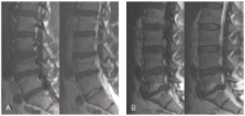
          Modic changes are bone marrow lesions visible in magnetic resonance imaging (MRI), and they are assumed to be associated with symptomatic intervertebral disc disease, especially changes located at L5-S1. Only limited information exists about the determinants of Modic changes. The objective of this study was to evaluate the determinants of vertebral endplate (Modic) changes, and whether they are similar for Modic changes and severe disc degeneration focusing on L5-S1 level.

          Methods

          228 middle-aged male workers (159 train engineers and 69 sedentary factory workers) from northern Finland underwent sagittal T1- and T2-weighted MRI. Modic changes and disc degeneration were analyzed from the scans. The participants responded to a questionnaire including items of occupational history and lifestyle factors. Logistic regression analysis was used to evaluate the associations between selected determinants (age, lifetime exercise, weight-related factors, fat percentage, smoking, alcohol use, lifetime whole-body vibration) and Modic type I and II changes, and severe disc degeneration (= grade V on Pfirrmann's classification).

          Results

          The prevalences of the Modic changes and severe disc degeneration were similar in the occupational groups. Age was significantly associated with all degenerative changes. In the age-adjusted analyses, only weight-related determinants (BMI, waist circumference) were associated with type II changes. Exposure to whole-body vibration, besides age, was the only significant determinant for severe disc degeneration. In the multivariate model, BMI was associated with type II changes at L5-S1 (OR 2.75 per one SD = 3 unit increment in BMI), and vibration exposure with severe disc degeneration at L5-S1 (OR 1.08 per one SD = 11-year increment in vibration exposure).

          Conclusion

          Besides age, weight-related factors seem important in the pathogenesis of Modic changes, whereas whole-body vibration was the only significant determinant of severe disc degeneration.

          Related collections

          Most cited references26

          • Record: found
          • Abstract: not found
          • Article: not found

          Imaging of degenerative disk disease.

            Bookmark
            • Record: found
            • Abstract: found
            • Article: not found

            Disc degeneration of the lumbar spine in relation to overweight.

            To study the association between overweight and lumbar disc degeneration. Population-based 4-y follow-up magnetic resonance imaging (MRI) study. The subjects were 129 working middle-aged men selected to the baseline magnetic resonance imaging (MRI) study from a cohort of 1832 men representing three occupations: machine drivers, construction carpenters, and office workers. The selection was based on the paticipants' age (40-45 y) and place of residence. MR images of the lumbar spines were obtained at baseline and at 4-y follow-up. Signal intensity of the nucleus pulposus of the discs L2/L3-L4/L5 was visually assessed by two readers using the adjacent cerebrospinal fluid as an intensity reference. The weight (at age 25 and 40-45 y) and height of the subjects, history of car driving, smoking, and back injuries were assessed by questionnaire. Multiple regression analyses allowing for occupation, history of car driving, smoking, and back injuries showed that persistent overweight (body mass index (BMI) > or =25 kg/m(2) at both ages) associated strongly with an increased risk of the number of lumbar discs with decreased signal intensity of nucleus pulposus at follow-up, adjusted odds ratio (OR) being 4.3 (95% confidence intervals (95% CIs) 1.3-14.3). Overweight at young age (risk ratio (RR) 3.8; 95% CI 1.4-10.4) was a stronger predictor of an increase in the number of degenerated discs during follow-up than overweight in middle age (RR 1.3; 95% CI 0.7-2.7). The study provides evidence that the BMI above 25 kg/m(2) increases the risk of lumbar disc degeneration. Overweight at young age seems to be particularly detrimental.
              Bookmark
              • Record: found
              • Abstract: found
              • Article: not found

              Magnetic resonance imaging and low back pain in adults: a diagnostic imaging study of 40-year-old men and women.

              Cross-sectional cohort study of a general population. To investigate "abnormal" lumbar spine magnetic resonance imaging (MRI) findings, and their prevalence and associations with low back pain (LBP). The clinical relevance of various "abnormal" findings in the lumbar spine is unclear. Distinguishing between inevitable age-related findings and degenerative findings with deleterious consequences is a challenge. Lumbar spine MRI was obtained in 412, 40-year-old individuals. Predefined "abnormal" MRI findings were interpreted without any knowledge of patient symptoms. Associations between MRI abnormalities and LBP were calculated using odds ratios. The "overall picture" of each MRI finding was established on the basis of the frequencies, diagnostic values, and the strength and consistency of associations. Most "abnormal" MRI findings were found at the lowest lumbar levels. Irregular nucleus shape and reduced disc height were common (>50% of individuals). Relatively common (25% to 50%) were hypointense disc signal, anular tears, high intensity zones, disc protrusions, endplate changes, zygapophyseal joint degeneration, asymmetry, and foraminal stenosis. Nerve root compromise, Modic changes, central spinal stenosis, and anterolisthesis/retrolisthesis were rare ( 4). Significantly positive associations with all LBP variables were seen for hypointense disc signals, reduced disc height, and Modic changes. All disc "abnormalities" except protrusion were moderately associated with LBP during the past year. Most degenerative disc "abnormalities" were moderately associated with LBP. The strongest associations were noted for Modic changes and anterolisthesis. Further studies are needed to define clinical relevance.
                Bookmark

                Author and article information

                Journal
                BMC Musculoskelet Disord
                BMC Musculoskeletal Disorders
                BioMed Central
                1471-2474
                2008
                16 April 2008
                : 9
                : 51
                Affiliations
                [1 ]Department of Diagnostic Radiology, Oulu University Hospital, Oulu, Finland
                [2 ]Department of Physical and Rehabilitation Medicine, Oulu University Hospital, Oulu, Finland
                [3 ]Public Health Institute, Helsinki, Finland
                [4 ]Department of Sports Medicine, Oulu Deaconess Institute, Oulu, Finland
                [5 ]Department of Public Health, University of Helsinki, Finland
                [6 ]Department of Orthopaedics, Tampere University Hospital, Tampere, Finland
                [7 ]Musculoskeletal Centre, Finnish Institute of Occupational Health, Oulu, Finland
                [8 ]ORTON Orthopedic Hospital, Helsinki, Finland
                [9 ]Department of Public Health and General Practice, University of Oulu, Finland
                Article
                1471-2474-9-51
                10.1186/1471-2474-9-51
                2373785
                18416819
                5105e152-8cea-46da-a552-be173c263ae4
                Copyright © 2008 Kuisma et al; licensee BioMed Central Ltd.

                This is an Open Access article distributed under the terms of the Creative Commons Attribution License ( http://creativecommons.org/licenses/by/2.0), which permits unrestricted use, distribution, and reproduction in any medium, provided the original work is properly cited.

                History
                : 18 October 2007
                : 16 April 2008
                Categories
                Research Article

                Orthopedics
                Orthopedics

                Comments

                Comment on this article