6
views
0
recommends
+1 Recommend
0 collections
    0
    shares
      • Record: found
      • Abstract: found
      • Article: found
      Is Open Access

      Aging of the skeletal muscle extracellular matrix drives a stem cell fibrogenic conversion

      research-article

      Read this article at

      Bookmark
          There is no author summary for this article yet. Authors can add summaries to their articles on ScienceOpen to make them more accessible to a non-specialist audience.

          Summary

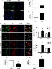
          Age‐related declines in skeletal muscle regeneration have been attributed to muscle stem cell (MuSC) dysfunction. Aged MuSCs display a fibrogenic conversion, leading to fibrosis and impaired recovery after injury. Although studies have demonstrated the influence of in vitro substrate characteristics on stem cell fate, whether and how aging of the extracellular matrix (ECM) affects stem cell behavior has not been investigated. Here, we investigated the direct effect of the aged muscle ECM on MuSC lineage specification. Quantification of ECM topology and muscle mechanical properties reveals decreased collagen tortuosity and muscle stiffening with increasing age. Age‐related ECM alterations directly disrupt MuSC responses, and MuSCs seeded ex vivo onto decellularized ECM constructs derived from aged muscle display increased expression of fibrogenic markers and decreased myogenicity, compared to MuSCs seeded onto young ECM. This fibrogenic conversion is recapitulated in vitro when MuSCs are seeded directly onto matrices elaborated by aged fibroblasts. When compared to young fibroblasts, fibroblasts isolated from aged muscle display increased nuclear levels of the mechanosensors, Yes‐associated protein (YAP)/transcriptional coactivator with PDZ‐binding motif (TAZ), consistent with exposure to a stiff microenvironment in vivo. Accordingly, preconditioning of young fibroblasts by seeding them onto a substrate engineered to mimic the stiffness of aged muscle increases YAP/TAZ nuclear translocation and promotes secretion of a matrix that favors MuSC fibrogenesis. The findings here suggest that an age‐related increase in muscle stiffness drives YAP/TAZ‐mediated pathogenic expression of matricellular proteins by fibroblasts, ultimately disrupting MuSC fate.

          Related collections

          Most cited references39

          • Record: found
          • Abstract: found
          • Article: not found

          Cellular mechanotransduction: putting all the pieces together again.

          Analysis of cellular mechanotransduction, the mechanism by which cells convert mechanical signals into biochemical responses, has focused on identification of critical mechanosensitive molecules and cellular components. Stretch-activated ion channels, caveolae, integrins, cadherins, growth factor receptors, myosin motors, cytoskeletal filaments, nuclei, extracellular matrix, and numerous other structures and signaling molecules have all been shown to contribute to the mechanotransduction response. However, little is known about how these different molecules function within the structural context of living cells, tissues, and organs to produce the orchestrated cellular behaviors required for mechanosensation, embryogenesis, and physiological control. Recent work from a wide range of fields reveals that organ, tissue, and cell anatomy are as important for mechanotransduction as individual mechanosensitive proteins and that our bodies use structural hierarchies (systems within systems) composed of interconnected networks that span from the macroscale to the nanoscale in order to focus stresses on specific mechanotransducer molecules. The presence of isometric tension (prestress) at all levels of these multiscale networks ensures that various molecular scale mechanochemical transduction mechanisms proceed simultaneously and produce a concerted response. Future research in this area will therefore require analysis, understanding, and modeling of tensionally integrated (tensegrity) systems of mechanochemical control.
            Bookmark
            • Record: found
            • Abstract: found
            • Article: not found

            Notch-mediated restoration of regenerative potential to aged muscle.

            A hallmark of aging is diminished regenerative potential of tissues, but the mechanism of this decline is unknown. Analysis of injured muscle revealed that, with age, resident precursor cells (satellite cells) had a markedly impaired propensity to proliferate and to produce myoblasts necessary for muscle regeneration. This was due to insufficient up-regulation of the Notch ligand Delta and, thus, diminished activation of Notch in aged, regenerating muscle. Inhibition of Notch impaired regeneration of young muscle, whereas forced activation of Notch restored regenerative potential to old muscle. Thus, Notch signaling is a key determinant of muscle regenerative potential that declines with age.
              Bookmark
              • Record: found
              • Abstract: found
              • Article: not found

              Connective tissue fibroblasts and Tcf4 regulate myogenesis.

              Muscle and its connective tissue are intimately linked in the embryo and in the adult, suggesting that interactions between these tissues are crucial for their development. However, the study of muscle connective tissue has been hindered by the lack of molecular markers and genetic reagents to label connective tissue fibroblasts. Here, we show that the transcription factor Tcf4 (transcription factor 7-like 2; Tcf7l2) is strongly expressed in connective tissue fibroblasts and that Tcf4(GFPCre) mice allow genetic manipulation of these fibroblasts. Using this new reagent, we find that connective tissue fibroblasts critically regulate two aspects of myogenesis: muscle fiber type development and maturation. Fibroblasts promote (via Tcf4-dependent signals) slow myogenesis by stimulating the expression of slow myosin heavy chain. Also, fibroblasts promote the switch from fetal to adult muscle by repressing (via Tcf4-dependent signals) the expression of developmental embryonic myosin and promoting (via a Tcf4-independent mechanism) the formation of large multinucleate myofibers. In addition, our analysis of Tcf4 function unexpectedly reveals a novel mechanism of intrinsic regulation of muscle fiber type development. Unlike other intrinsic regulators of fiber type, low levels of Tcf4 in myogenic cells promote both slow and fast myogenesis, thereby promoting overall maturation of muscle fiber type. Thus, we have identified novel extrinsic and intrinsic mechanisms regulating myogenesis. Most significantly, our data demonstrate for the first time that connective tissue is important not only for adult muscle structure and function, but is a vital component of the niche within which muscle progenitors reside and is a critical regulator of myogenesis.
                Bookmark

                Author and article information

                Contributors
                ambrosiof@upmc.edu
                Journal
                Aging Cell
                Aging Cell
                10.1111/(ISSN)1474-9726
                ACEL
                Aging Cell
                John Wiley and Sons Inc. (Hoboken )
                1474-9718
                1474-9726
                30 March 2017
                June 2017
                : 16
                : 3 ( doiID: 10.1111/acel.2017.16.issue-3 )
                : 518-528
                Affiliations
                [ 1 ] Department of Physical Medicine and RehabilitationUniversity of Pittsburgh Kaufmann Medical Building, Suite 201, 3471 Fifth Avenue Pittsburgh PA 15213USA
                [ 2 ] McGowan Institute for Regenerative MedicineUniversity of Pittsburgh 450 Technology Drive, Suite 300 Pittsburgh PA 15219USA
                [ 3 ] Department of SurgeryUniversity of Pittsburgh 450 Technology Drive, Suite 300 Pittsburgh PA 15219USA
                [ 4 ] Department of Environmental and Occupational HealthUniversity of Pittsburgh 100 Technology Drive, Suite 328 Pittsburgh PA 15219USA
                [ 5 ] Center for Cellular and Molecular Engineering Department of Orthopaedic SurgeryUniversity of Pittsburgh 450 Technology Drive, Bridgeside Point II, Suite 221 Pittsburgh PA 15219USA
                [ 6 ] Center for Vascular Remodeling and Regeneration Center for Bioengineering (CNBIO)University of Pittsburgh 300 Technology Drive, Suite 300 Pittsburgh PA 15219USA
                [ 7 ] Department of BioengineeringUniversity of Pittsburgh 213 Center for Bioengineering, 300 Technology Drive Pittsburgh PA 15219USA
                [ 8 ] Department of EngineeringUniversity of Leicester 127 Michael Atiyah Building, University Road Leicester LE1 7RHUK
                [ 9 ] Glenn Center for the Biology of Aging and Department of Neurology and Neurological Sciences Stanford University School of Medicine Stanford CA 94305USA
                [ 10 ] RR&D CenterVA Palo Alto Health Care System Palo Alto CA 94304USA
                Author notes
                [*] [* ] Correspondence

                Fabrisia Ambrosio, PhD, MPT, 450 Technology Drive, Bridgeside Point II, Suite 308, Pittsburgh, PA 15219, USA. Tel.: +412 624 5276; fax: +412‐624‐5260; e‐mail : ambrosiof@ 123456upmc.edu

                Article
                ACEL12578
                10.1111/acel.12578
                5418187
                28371268
                507739a0-db3e-4f5f-aad7-4e42755a397c
                © 2017 The Authors. Aging Cell published by the Anatomical Society and John Wiley & Sons Ltd.

                This is an open access article under the terms of the Creative Commons Attribution License, which permits use, distribution and reproduction in any medium, provided the original work is properly cited.

                History
                : 08 January 2017
                Page count
                Figures: 6, Tables: 0, Pages: 11, Words: 8513
                Funding
                Funded by: NIH
                Funded by: NIA
                Award ID: K01AG039477
                Funded by: NIEHS
                Award ID: F32ES022134
                Award ID: R01ES023696
                Award ID: R01ES025529
                Funded by: University of Pittsburgh Medical Center Rehabilitation Institute
                Categories
                Original Article
                Original Articles
                Custom metadata
                2.0
                acel12578
                June 2017
                Converter:WILEY_ML3GV2_TO_NLMPMC version:5.0.9 mode:remove_FC converted:18.05.2017

                Cell biology
                aging,extracellular matrix,satellite cells,muscle stem cells,skeletal muscle
                Cell biology
                aging, extracellular matrix, satellite cells, muscle stem cells, skeletal muscle

                Comments

                Comment on this article