7
views
0
recommends
+1 Recommend
0 collections
    0
    shares
      • Record: found
      • Abstract: found
      • Article: found
      Is Open Access

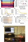
      Advancements in Dermatological Imaging Modalities

      other

      Read this article at

      Bookmark
          There is no author summary for this article yet. Authors can add summaries to their articles on ScienceOpen to make them more accessible to a non-specialist audience.

          Related collections

          Most cited references93

          • Record: found
          • Abstract: found
          • Article: not found

          Medical applications of infrared thermography: A review

          Abnormal body temperature is a natural indicator of illness. Infrared thermography (IRT) is a fast, passive, non-contact and non-invasive alternative to conventional clinical thermometers for monitoring body temperature. Besides, IRT can also map body surface temperature remotely. Last five decades witnessed a steady increase in the utility of thermal imaging cameras to obtain correlations between the thermal physiology and skin temperature. IRT has been successfully used in diagnosis of breast cancer, diabetes neuropathy and peripheral vascular disorders. It has also been used to detect problems associated with gynecology, kidney transplantation, dermatology, heart, neonatal physiology, fever screening and brain imaging. With the advent of modern infrared cameras, data acquisition and processing techniques, it is now possible to have real time high resolution thermographic images, which is likely to surge further research in this field. The present efforts are focused on automatic analysis of temperature distribution of regions of interest and their statistical analysis for detection of abnormalities. This critical review focuses on advances in the area of medical IRT. The basics of IRT, essential theoretical background, the procedures adopted for various measurements and applications of IRT in various medical fields are discussed in this review. Besides background information is provided for beginners for better understanding of the subject.
            Bookmark
            • Record: found
            • Abstract: found
            • Article: found
            Is Open Access

            A review of clinical photoacoustic imaging: Current and future trends

            Photoacoustic imaging (or optoacoustic imaging) is an upcoming biomedical imaging modality availing the benefits of optical resolution and acoustic depth of penetration. With its capacity to offer structural, functional, molecular and kinetic information making use of either endogenous contrast agents like hemoglobin, lipid, melanin and water or a variety of exogenous contrast agents or both, PAI has demonstrated promising potential in a wide range of preclinical and clinical applications. This review provides an overview of the rapidly expanding clinical applications of photoacoustic imaging including breast imaging, dermatologic imaging, vascular imaging, carotid artery imaging, musculoskeletal imaging, gastrointestinal imaging and adipose tissue imaging and the future directives utilizing different configurations of photoacoustic imaging. Particular emphasis is placed on investigations performed on human or human specimens.
              Bookmark
              • Record: found
              • Abstract: found
              • Article: not found

              Optical coherence tomography in dermatology: a review.

              J Welzel (2001)
              Optical coherence tomography (OCT) is a non-invasive technique for morphological investigation of tissue. Since its development in the late 1980s it is mainly used as a diagnostic tool in ophthalmology. For examination of a highly scattering tissue like the skin, it was necessary to modify the method. Early studies on the value of OCT for skin diagnosis gave promising results. The OCT technique is based on the principle of Michelson interferometry. The light sources used for OCT are low coherent superluminescent diodes operating at a wavelength of about 1300 nm. OCT provides two-dimensional images with a scan length of a few millimeters (mm), a resolution of about 15 microns and a maximum detection depth of 1.5 mm. The image acquisition can be performed nearly in real time. The measurement is non-invasive and with no side effects. The in vivo OCT images of human skin show a strong scattering from tissue with a few layers and some optical inhomogeneities. The resolution enables the visualization of architectural changes, but not of single cells. In palmoplantar skin, the thick stratum comeum is visible as a low-scattering superficial well defined layer with spiral sweat gland ducts inside. The epidermis can be distinguished from the dermis. Adnexal structures and blood vessels are low-scattering regions in the upper dermis. Skin tumors show a homogenous signal distribution. In some cases, tumor borders to healthy skin are detectable. Inflammatory skin diseases lead to changes of the OCT image, such as thickening of the epidermis and reduction of the light attenuation in the dermis. A quantification of treatment effects, such as swelling of the horny layer due to application of a moisturizer, is possible. Repeated measurements allow a monitoring of the changes over time. OCT is a promising new bioengineering method for investigation of skin morphology. In some cases it may be useful for diagnosis of skin diseases. Because of its non-invasive character, the technique allows monitoring of inflammatory diseases over time. An objective quantification of the efficacy and tolerance of topical treatment is also possible. Due to the high resolution and simple application, OCT is an interesting addition to other morphological techniques in dermatology.
                Bookmark

                Author and article information

                Journal
                Indian Dermatol Online J
                Indian Dermatol Online J
                IDOJ
                Indian Dermatol Online J
                Indian Dermatology Online Journal
                Wolters Kluwer - Medknow (India )
                2229-5178
                2249-5673
                Mar-Apr 2024
                28 February 2024
                : 15
                : 2
                : 278-292
                Affiliations
                [1] Department of Dermatology and Venereology, All India Institute of Medical Sciences, New Delhi, Delhi, India
                [1 ] Financial Research and Executive Insights, Everest Group, Gurugram, Haryana, India
                Author notes
                Address for correspondence: Dr. Somesh Gupta, Department of Dermatology and Venereology, All India Institute of Medical Sciences, New Delhi, Delhi - 110 029, India. E-mail: someshgupta@ 123456aiims.edu
                Article
                IDOJ-15-278
                10.4103/idoj.idoj_852_23
                10969257
                38550821
                5039d015-fe2c-4f76-bfc8-243976993c2b
                Copyright: © 2024 Indian Dermatology Online Journal

                This is an open access journal, and articles are distributed under the terms of the Creative Commons Attribution-NonCommercial-ShareAlike 4.0 License, which allows others to remix, tweak, and build upon the work non-commercially, as long as appropriate credit is given and the new creations are licensed under the identical terms.

                History
                : 14 November 2023
                : 29 December 2023
                : 25 December 2023
                Categories
                Resident’s Area

                Dermatology
                Dermatology

                Comments

                Comment on this article