3
views
0
recommends
+1 Recommend
0 collections
    0
    shares
      • Record: found
      • Abstract: found
      • Article: found
      Is Open Access

      Anterior debridement, bone grafting and fixation for cervical spine tuberculosis: an iliac bone graft versus a structural manubrium graft

      research-article

      Read this article at

      Bookmark
          There is no author summary for this article yet. Authors can add summaries to their articles on ScienceOpen to make them more accessible to a non-specialist audience.

          Abstract

          Background

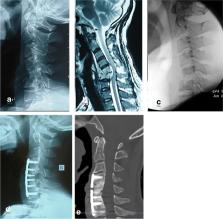
          Anterior debridement, decompression, bone grafting, and instrumentation are safe and effective techniques for patients with lower cervical spine tuberculosis. However, there is no consensus regarding the methods for using autogenous bone grafts. The purpose of this retrospective study was to compare the clinical outcomes of anterior surgical management for cervical spine tuberculosis by using an iliac bone graft versus a structural manubrium graft.

          Methods

          From January 2009 to September 2018, 23 patients with cervical spine tuberculosis were treated with anterior debridement, autogenous structural bone grafting and fixation at our spinal department. The patients were divided into 2 groups according to the different graft materials, namely, iliac crest bone grafts (Group A) and structural manubrium grafts (Group B). The clinical and radiographic results of the 2 groups were analyzed and compared.

          Results

          The mean duration of follow-up was 24 months. Bony fusion was achieved in all patients without failure of internal fixation. There were no significant differences between the two groups with respect to the operation time, blood loss, fusion time, neurological outcomes, or postoperative local Cobb angle ( P > .05). However, the donor site complication rate in Group A was greater than that in Group B. The postoperative ambulation time in Group A was later than that in Group B. The mean visual analog scale (VAS) score for donor site pain in Group A was higher than that in Group B at 1 week after surgery ( P < 0.05). However, there was no significant difference between the 2 groups at the last visit ( P > .05).

          Conclusion

          Both iliac bone grafts and sternal manubrium grafts can effectively reconstruct anterior column defects in anterior surgery. However, structural sternal manubrium autografts cause fewer complications associated with donor site morbidities than iliac bone grafts.

          Related collections

          Most cited references15

          • Record: found
          • Abstract: found
          • Article: not found

          Autogenous bone graft: donor sites and techniques.

          Autogenous cancellous bone graft provides an osteoconductive, osteoinductive, and osteogenic substrate for filling bone voids and augmenting fracture-healing.The iliac crest remains the most frequently used site for bone-graft harvest, but the proximal part of the tibia, distal end of the radius, distal aspect of the tibia, and greater trochanter are alternative donor sites that are particularly useful for bone-grafting in the ipsilateral extremity.The most common complication associated with the harvest of autogenous bone graft is pain at the donor site, with less frequent complications including nerve injury, hematoma, infection, and fracture at the donor site.Induced membranes is a method that uses a temporary polymethylmethacrylate cement spacer to create a bone-graft-friendly environment to facilitate graft incorporation, even in large segmental defects.
            Bookmark
            • Record: found
            • Abstract: found
            • Article: not found

            Preliminary experience using a polyetheretherketone (PEEK) cage in the treatment of cervical disc disease.

            We investigated the effectiveness of a new material, polyetheretherketone (PEEK), in a spinal cage used in performing cervical spinal fusion for the correction of cervical kyphosis. A total of 80 patients with various cervical diseases were divided into two groups. Patients in Group A (40 patients) underwent microdiscectomy and PEEK cage fusion, and patients in Group B (40 patients) were treated with microdisectomy and autogenous iliac crest graft (AICG) fusion. We evaluated the patients for cervical lordosis, the height of the foramina, the cross sectional area of the foramina, and fusion status on the basis of x-rays. The patients' neurological and functional outcomes were assessed on the basis of the Prolo scale. Magnetic resonance imaging was also performed for spinal cord evaluation. The use of the PEEK cage in patients who undergo spinal fusion may increase cervical lordosis (mean, 2.33 +/- 3.00 mm; P = 0.03), whereas AICG fusion may not (mean, -0.84 +/- 6.69 mm; P = 0.49). The use of the PEEK cage was found to increase the height of the foramina (mean, 2.54 +/- 1.40 mm; P = 0.00) and increase its cross sectional area (mean, 40.36 +/- 23.53 mm2; P = 0.00). The height of the foramina increased only in the PEEK group postoperatively. The cross sectional area of the foramina increased in both groups postoperatively. The complication rate in patients who underwent fusion procedures with the PEEK cage was less than that in patients who underwent fusion with AICG fusion (2.50 versus 17.50%; P = 0.03). Both groups had a satisfactory fusion rate (100 versus 93.1%). The patients' postoperative Prolo scale scores were statistically better in the PEEK group (8.50 +/- 1.49 versus 7.17 +/- 2.13; P = 0.00), and more patients in the PEEK group than in the AICG group achieved excellent outcomes (66.63 versus 28.57%; P = 0.00). Because PEEK is radiotransparent on x-rays and few artifacts are seen on magnetic resonance imaging scans, it is better suited than autogenous iliac crest donor material for postoperative radiographic evaluation. The PEEK cage provides solid fusion, increased cervical lordosis, and increased height and cross sectional area of the foramina. There are few complications associated with the use of this cage, and the functional and neurological outcomes are satisfactory. It also facilitates postoperative x-ray and magnetic resonance imaging evaluation. The PEEK cage is therefore a good substitute for AICG fusion in patients with cervical disc disease.
              Bookmark
              • Record: found
              • Abstract: found
              • Article: found
              Is Open Access

              Anterior versus posterior procedure for surgical treatment of thoracolumbar tuberculosis: A retrospective analysis

              Background: Approach for surgical treatment of thoracolumbar tuberculosis has been controversial. The aim of present study is to compare the clinical, radiological and functional outcome of anterior versus posterior debridement and spinal fixation for the surgical treatment of thoracic and thoracolumbar tuberculosis. Materials and Methods: 70 patients with spinal tuberculosis treated surgically between Jan 2001 and Dec 2006 were included in the study. Thirty four patients (group I) with mean age 34.9 years underwent anterior debridement, decompression and instrumentation by anterior transthoracic, transpleural and/or retroperitoneal diaphragm cutting approach. Thirty six patients (group II) with mean age of 33.6 years were operated by posterolateral (extracavitary) decompression and posterior instrumentation. Various parameters like blood loss, surgical time, levels of instrumentation, neurological recovery, and kyphosis improvement were compared. Fusion assessment was done as per Bridwell criteria. Functional outcome was assessed using Prolo scale. Mean followup was 26 months. Results: Mean surgical time in group I was 5 h 10 min versus 4 h 50 min in group II (P>0.05). Average blood loss in group I was 900 ml compared to 1100 ml in group II (P>0.05). In group I, the percentage immediate correction in kyphosis was 52.27% versus 72.80% in group II. Satisfactory bony fusion (grades I and II) was seen in 100% patients in group I versus 97.22% in group II. Three patients in group I needed prolonged immediate postoperative ICU support compared to one in group II. Injury to lung parenchyma was seen in one patient in group I while the anterior procedure had to be abandoned in one case due to pleural adhesions. Functional outcome (Prolo scale) in group II was good in 94.4% patients compared to 88.23% patients in group I. Conclusion: Though the anterior approach is an equally good method for debridement and stabilization, kyphus correction is better with posterior instrumentation and the posterior approach is associated with less morbidity and complications.
                Bookmark

                Author and article information

                Contributors
                hx_sym@163.com
                wqspine2004@163.com
                Journal
                BMC Musculoskelet Disord
                BMC Musculoskelet Disord
                BMC Musculoskeletal Disorders
                BioMed Central (London )
                1471-2474
                11 March 2022
                11 March 2022
                2022
                : 23
                : 236
                Affiliations
                [1 ]GRID grid.412901.f, ISNI 0000 0004 1770 1022, Department of Orthopedics, , Orthopedic Research Institute, West China Hospital, Sichuan University, ; Chengdu, 646000 Sichuan China
                [2 ]GRID grid.488387.8, Department of Orthopedic Surgery, , Affiliated Hospital of Southwest Medical University, ; No. 25 of Taiping Road, Luzhou, 646000 Sichuan China
                Article
                5177
                10.1186/s12891-022-05177-0
                8915494
                35277155
                5031a032-aa87-4dc6-8015-5e28473f48da
                © The Author(s) 2022

                Open AccessThis article is licensed under a Creative Commons Attribution 4.0 International License, which permits use, sharing, adaptation, distribution and reproduction in any medium or format, as long as you give appropriate credit to the original author(s) and the source, provide a link to the Creative Commons licence, and indicate if changes were made. The images or other third party material in this article are included in the article's Creative Commons licence, unless indicated otherwise in a credit line to the material. If material is not included in the article's Creative Commons licence and your intended use is not permitted by statutory regulation or exceeds the permitted use, you will need to obtain permission directly from the copyright holder. To view a copy of this licence, visit http://creativecommons.org/licenses/by/4.0/. The Creative Commons Public Domain Dedication waiver ( http://creativecommons.org/publicdomain/zero/1.0/) applies to the data made available in this article, unless otherwise stated in a credit line to the data.

                History
                : 25 May 2021
                : 3 March 2022
                Categories
                Research
                Custom metadata
                © The Author(s) 2022

                Orthopedics
                manubrium,autograft,cervical tuberculosis,kyphosis,instrumentation
                Orthopedics
                manubrium, autograft, cervical tuberculosis, kyphosis, instrumentation

                Comments

                Comment on this article