13
views
0
recommends
+1 Recommend
0 collections
    0
    shares
      • Record: found
      • Abstract: found
      • Article: not found

      Two Distinct Pathways Mediated by PA28 and hsp90 in Major Histocompatibility Complex Class I Antigen Processing

      research-article

      Read this article at

      ScienceOpenPublisherPMC
      Bookmark
          There is no author summary for this article yet. Authors can add summaries to their articles on ScienceOpen to make them more accessible to a non-specialist audience.

          Abstract

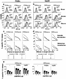
          Major histocompatibility complex (MHC) class I ligands are mainly produced by the proteasome. Herein, we show that the processing of antigens is regulated by two distinct pathways, one requiring PA28 and the other hsp90. Both hsp90 and PA28 enhanced the antigen processing of ovalbumin (OVA). Geldanamycin, an inhibitor of hsp90, almost completely suppressed OVA antigen presentation in PA28α −/−−/− lipopolysaccharide blasts, but not in wild-type cells, indicating that hsp90 compensates for the loss of PA28 and is essential in the PA28-independent pathway. In contrast, treatment of cells with interferon (IFN)-γ, which induces PA28 expression, abrogated the requirement of hsp90, suggesting that IFN-γ enhances the PA28-dependent pathway, whereas it diminishes hsp90-dependent pathway. Importantly, IFN-γ did not induce MHC class I expressions in PA28-deficient cells, indicating a prominent role for PA28 in IFN-γ–stimulated peptide supply. Thus, these two pathways operate either redundantly or specifically, depending on antigen species and cell type.

          Related collections

          Most cited references34

          • Record: found
          • Abstract: found
          • Article: not found

          The 26S proteasome: a molecular machine designed for controlled proteolysis.

          In eukaryotic cells, most proteins in the cytosol and nucleus are degraded via the ubiquitin-proteasome pathway. The 26S proteasome is a 2.5-MDa molecular machine built from approximately 31 different subunits, which catalyzes protein degradation. It contains a barrel-shaped proteolytic core complex (the 20S proteasome), capped at one or both ends by 19S regulatory complexes, which recognize ubiquitinated proteins. The regulatory complexes are also implicated in unfolding and translocation of ubiquitinated targets into the interior of the 20S complex, where they are degraded to oligopeptides. Structure, assembly and enzymatic mechanism of the 20S complex have been elucidated, but the functional organization of the 19S complex is less well understood. Most subunits of the 19S complex have been identified, however, specific functions have been assigned to only a few. A low-resolution structure of the 26S proteasome has been obtained by electron microscopy, but the precise arrangement of subunits in the 19S complex is unclear.
            Bookmark
            • Record: found
            • Abstract: found
            • Article: not found

            Identification and structural characterization of the ATP/ADP-binding site in the Hsp90 molecular chaperone.

            Hsp90 molecular chaperones in eukaryotic cells play essential roles in the folding and activation of a range of client proteins involved in cell cycle regulation, steroid hormone responsiveness, and signal transduction. The biochemical mechanism of Hsp90 is poorly understood, and the involvement of ATP in particular is controversial. Crystal structures of complexes between the N-terminal domain of the yeast Hsp90 chaperone and ADP/ATP unambiguously identify a specific adenine nucleotide binding site homologous to the ATP-binding site of DNA gyrase B. This site is the same as that identified for the antitumor agent geldanamycin, suggesting that geldanamycin acts by blocking the binding of nucleotides to Hsp90 and not the binding of incompletely folded client polypeptides as previously suggested. These results finally resolve the question of the direct involvement of ATP in Hsp90 function.
              Bookmark
              • Record: found
              • Abstract: found
              • Article: not found

              The co-chaperone CHIP regulates protein triage decisions mediated by heat-shock proteins.

              To maintain quality control in cells, mechanisms distinguish among improperly folded peptides, mature and functional proteins, and proteins to be targeted for degradation. The molecular chaperones, including heat-shock protein Hsp90, have the ability to recognize misfolded proteins and assist in their conversion to a functional conformation. Disruption of Hsp90 heterocomplexes by the Hsp90 inhibitor geldanamycin leads to substrate degradation through the ubiquitin-proteasome pathway, implicating this system in protein triage decisions. We previously identified CHIP (carboxyl terminus of Hsc70-interacting protein) to be an interaction partner of Hsc70 (ref. 4). CHIP also interacts directly with a tetratricopeptide repeat acceptor site of Hsp90, incorporating into Hsp90 heterocomplexes and eliciting release of the regulatory cofactor p23. Here we show that CHIP abolishes the steroid-binding activity and transactivation potential of the glucocorticoid receptor, a well-characterized Hsp90 substrate, even though it has little effect on its synthesis. Instead, CHIP induces ubiquitylation of the glucocorticoid receptor and degradation through the proteasome. By remodelling Hsp90 heterocomplexes to favour substrate degradation, CHIP modulates protein triage decisions that regulate the balance between protein folding and degradation for chaperone substrates.
                Bookmark

                Author and article information

                Journal
                J Exp Med
                The Journal of Experimental Medicine
                The Rockefeller University Press
                0022-1007
                1540-9538
                15 July 2002
                : 196
                : 2
                : 185-196
                Affiliations
                [1 ]Department of Molecular Medicine, Division of Immunology, Nagasaki University School of Medicine, Nagasaki 852-8523, Japan
                [2 ]Department of Molecular Oncology, Tokyo Metropolitan Institute of Medical Science, and CREST, Japan Science and Technology Corporation, Tokyo 113-8613, Japan
                [3 ]UpScience, Incorporated, Yokohama 244-8588, Japan
                [4 ]First Department of Surgery, Okayama University Medical School, Okayama 700, Japan
                Author notes

                Address correspondence to Heiichiro Udono, Department of Molecular Medicine, Division of Immunology, Nagasaki University School of Medicine, Nagasaki 852-8523, Japan. Phone: 81-95-849-7071; Fax: 81-95-849-7073; E-mail: udonoh@ 123456net.nagasaki-u.ac.jp

                Article
                20011922
                10.1084/jem.20011922
                2193925
                12119343
                502d28d0-112f-4b4a-819b-bb682a56dbe6
                Copyright © 2002, The Rockefeller University Press
                History
                : 16 November 2001
                : 10 May 2002
                : 3 June 2002
                Categories
                Article

                Medicine
                antigen presentation,transplantation immunology,immunity active,macrophage activation,cytotoxic t lymphocytes

                Comments

                Comment on this article