5
views
0
recommends
+1 Recommend
0 collections
    0
    shares
      • Record: found
      • Abstract: found
      • Article: found
      Is Open Access

      Chemotherapeutic Stress Influences Epithelial–Mesenchymal Transition and Stemness in Cancer Stem Cells of Triple-Negative Breast Cancer

      research-article

      Read this article at

      Bookmark
          There is no author summary for this article yet. Authors can add summaries to their articles on ScienceOpen to make them more accessible to a non-specialist audience.

          Abstract

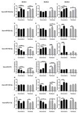
          Triple-negative breast cancer (TNBC) is a subtype of breast cancer characterized by the absence of estrogen and progesterone receptors (ER, PR) and lacking an overexpression of human epidermal growth factor receptor 2 (HER2). Apart from this lack of therapeutic targets, TNBC also shows an increased capacity for early metastasis and therapy resistance. Currently, many TNBC patients receive neoadjuvant chemotherapy (NACT) upon detection of the disease. With TNBC likely being driven at least in part by a cancer stem-like cell type, we wanted to evaluate the response of primary cancer stem cells (CSCs) to standard chemotherapeutics. Therefore, we set up a survival model using primary CSCs to mimic tumor cells in patients under chemotherapy. Breast cancer stem cells (BCSCs) were exposed to chemotherapeutics with a sublethal dose for six days. Surviving cells were allowed to recover in culture medium without chemotherapeutics. Surviving and recovered cells were examined in regard to proliferation, migratory capacity, sphere forming capacity, epithelial–mesenchymal transition (EMT) factor expression at the mRNA level, and cancer-related microRNA (miRNA) profile. Our results indicate that chemotherapeutic stress enhanced sphere forming capacity of BCSCs, and changed cell morphology and EMT-related gene expression at the mRNA level, whereas the migratory capacity was unaffected. Six miRNAs were identified as potential regulators in this process.

          Related collections

          Most cited references52

          • Record: found
          • Abstract: found
          • Article: found
          Is Open Access

          Human breast cancer cell lines contain stem-like cells that self-renew, give rise to phenotypically diverse progeny and survive chemotherapy

          Introduction The phenotypic and functional differences between cells that initiate human breast tumors (cancer stem cells) and those that comprise the tumor bulk are difficult to study using only primary tumor tissue. We embarked on this study hypothesizing that breast cancer cell lines would contain analogous hierarchical differentiation programs to those found in primary breast tumors. Methods Eight human breast cell lines (human mammary epithelial cells, and MCF10A, MCF7, SUM149, SUM159, SUM1315 and MDA.MB.231 cells) were analyzed using flow cytometry for CD44, CD24, and epithelial-specific antigen (ESA) expression. Limiting dilution orthotopic injections were used to evaluate tumor initiation, while serial colony-forming unit, reconstitution and tumorsphere assays were performed to assess self-renewal and differentiation. Pulse-chase bromodeoxyuridine (5-bromo-2-deoxyuridine [BrdU]) labeling was used to examine cell cycle and label-retention of cancer stem cells. Cells were treated with paclitaxol and 5-fluorouracil to test selective resistance to chemotherapy, and gene expression profile after chemotherapy were examined. Results The percentage of CD44+/CD24- cells within cell lines does not correlate with tumorigenicity, but as few as 100 cells can form tumors when sorted for CD44+/CD24-/low/ESA+. Furthermore, CD44+/CD24-/ESA+ cells can self-renew, reconstitute the parental cell line, retain BrdU label, and preferentially survive chemotherapy. Conclusion These data validate the use of cancer cell lines as models for the development and testing of novel therapeutics aimed at eradicating cancer stem cells.
            Bookmark
            • Record: found
            • Abstract: found
            • Article: not found

            XBP1 Promotes Triple Negative Breast Cancer By Controlling the HIF1 α Pathway

            Cancer cells induce a set of adaptive response pathways to survive in the face of stressors due to inadequate vascularization 1 . One such adaptive pathway is the unfolded protein (UPR) or endoplasmic reticulum (ER) stress response mediated in part by the ER-localized transmembrane sensor IRE1 2 and its substrate XBP1 3 . Previous studies report UPR activation in various human tumors 4-6 , but XBP1's role in cancer progression in mammary epithelial cells is largely unknown. Triple negative breast cancer (TNBC), a form of breast cancer in which tumor cells do not express the genes for estrogen receptor, progesterone receptor, and Her2/neu, is a highly aggressive malignancy with limited treatment options 7, 8 . Here, we report that XBP1 is activated in TNBC and plays a pivotal role in the tumorigenicity and progression of this human breast cancer subtype. In breast cancer cell line models, depletion of XBP1 inhibited tumor growth and tumor relapse and reduced the CD44high/CD24low population. Hypoxia-inducing factor (HIF)1α is known to be hyperactivated in TNBCs 9, 10 . Genome-wide mapping of the XBP1 transcriptional regulatory network revealed that XBP1 drives TNBC tumorigenicity by assembling a transcriptional complex with HIF1α that regulates the expression of HIF1α targets via the recruitment of RNA polymerase II. Analysis of independent cohorts of patients with TNBC revealed a specific XBP1 gene expression signature that was highly correlated with HIF1α and hypoxia-driven signatures and that strongly associated with poor prognosis. Our findings reveal a key function for the XBP1 branch of the UPR in TNBC and imply that targeting this pathway may offer alternative treatment strategies for this aggressive subtype of breast cancer.
              Bookmark
              • Record: found
              • Abstract: found
              • Article: found
              Is Open Access

              Mechanisms of Chemotherapy Resistance in Triple-Negative Breast Cancer—How We Can Rise to the Challenge

              Triple-negative (TNBC) is the most lethal subtype of breast cancer owing to high heterogeneity, aggressive nature, and lack of treatment options. Chemotherapy remains the standard of care for TNBC treatment, but unfortunately, patients frequently develop resistance. Accordingly, in recent years, tremendous effort has been made into elucidating the mechanisms of TNBC chemoresistance with the goal of identifying new molecular targets. It has become evident that the development of TNBC chemoresistance is multifaceted and based on the elaborate interplay of the tumor microenvironment, drug efflux, cancer stem cells, and bulk tumor cells. Alterations of multiple signaling pathways govern these interactions. Moreover, TNBC’s high heterogeneity, highlighted in the existence of several molecular signatures, presents a significant obstacle to successful treatment. In the present, in-depth review, we explore the contribution of key mechanisms to TNBC chemoresistance as well as emerging strategies to overcome them. We discuss novel anti-tumor agents that target the components of these mechanisms and pay special attention to their current clinical development while emphasizing the challenges still ahead of successful TNBC management. The evidence presented in this review outlines the role of crucial pathways in TNBC survival following chemotherapy treatment and highlights the importance of using combinatorial drug strategies and incorporating biomarkers in clinical studies.
                Bookmark

                Author and article information

                Journal
                Int J Mol Sci
                Int J Mol Sci
                ijms
                International Journal of Molecular Sciences
                MDPI
                1422-0067
                08 January 2020
                January 2020
                : 21
                : 2
                : 404
                Affiliations
                [1 ]Department of Obstetrics and Gynecology, University Hospital Aachen (UKA), 52074 Aachen, Germany; xli@ 123456ukaachen.de (X.L.); ableilevens@ 123456ukaachen.de (A.B.); estickeler@ 123456ukaachen.de (E.S.)
                [2 ]Department of Immunology, University of Freiburg, 79104 Freiburg, Germany; juliane.strietz@ 123456biologie.uni-freiburg.de
                Author notes
                [* ]Correspondence: jmaurer@ 123456ukaachen.de ; Tel.: +49-2418037051
                Author information
                https://orcid.org/0000-0001-9504-605X
                Article
                ijms-21-00404
                10.3390/ijms21020404
                7014166
                31936348
                4fe1b868-325a-4591-a5ec-251c6e2be5d3
                © 2020 by the authors.

                Licensee MDPI, Basel, Switzerland. This article is an open access article distributed under the terms and conditions of the Creative Commons Attribution (CC BY) license ( http://creativecommons.org/licenses/by/4.0/).

                History
                : 11 November 2019
                : 31 December 2019
                Categories
                Article

                Molecular biology
                breast cancer stem cell,chemotherapeutic stress,emt,stemness,mirna
                Molecular biology
                breast cancer stem cell, chemotherapeutic stress, emt, stemness, mirna

                Comments

                Comment on this article