4
views
0
recommends
+1 Recommend
0 collections
    0
    shares
      • Record: found
      • Abstract: found
      • Article: found
      Is Open Access

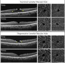
      Optical coherence tomography angiographic findings of lamellar macular hole: comparisons between tractional and degenerative subtypes

      research-article

      Read this article at

      Bookmark
          There is no author summary for this article yet. Authors can add summaries to their articles on ScienceOpen to make them more accessible to a non-specialist audience.

          Abstract

          We investigated the microvascular changes in eyes with lamellar macular holes (LMHs) using optical coherence tomography angiography (OCTA), compare them between two subtypes of LMH. Tractional and degenerative LMH were differentiated based on the morphological characteristics of OCT. In OCTA images, foveal and parafoveal vessel density (VD) in the superficial and deep capillary plexus (SCP, DCP) and foveal avascular zone (FAZ) area were measured. Eyes that underwent vitrectomy for LMH were included in subgroup analysis. We analysed 63 LMH (42 tractional and 21 degenerative) eyes and 63 control eyes. Compared with degenerative LMH, tractional LMH had better BCVA ( p = 0.010), smaller FAZ area ( p = 0.001), and higher foveal VD in the SCP ( p = 0.130) and DCP ( p = 0.002). In degenerative LMH, better BCVA was associated with greater foveal VD in the SCP ( p = 0.040) and DCP ( p = 0.005), and parafoveal VD in the SCP ( p = 0.006). In subgroup analysis, only the tractional LMH group showed significant increases in foveal and parafoveal VDs in the SCP after vitrectomy ( p = 0.001 and p = 0.026, respectively). Significant differences in microvascular changes were noted between tractional and degenerative LMH, suggesting that two subtypes are distinct pathogenetic entities.

          Related collections

          Most cited references23

          • Record: found
          • Abstract: found
          • Article: not found

          Redefining lamellar holes and the vitreomacular interface: an ultrahigh-resolution optical coherence tomography study.

          To define optical coherence tomographic (OCT) criteria for the diagnosis of a lamellar macular hole, and to increase understanding of lamellar hole pathogenesis by examining fine anatomic features using ultrahigh-resolution optical coherence tomography (UHR OCT). Retrospective observational case series. Nineteen eyes of 18 patients with lamellar holes were imaged with UHR OCT between 2002 and 2004. A UHR OCT system was developed for use in the ophthalmology clinic. All 6 UHR OCT images for each eye imaged were examined. Lamellar holes were diagnosed based on a characteristic OCT appearance. Criteria for the OCT diagnosis of a lamellar hole were as follows: (1) irregular foveal contour; (2) break in the inner fovea; (3) intraretinal split; and (4) intact foveal photoreceptors. From 1205 eyes of 664 patients imaged with UHR OCT, and retrospectively reviewed, 19 eyes of 18 patients were diagnosed with a lamellar hole based on these criteria. All 19 eyes were also imaged with standard resolution OCT. Their charts were retrospectively reviewed. Standard and ultrahigh-resolution OCT images. On chart review, clinical diagnosis of a lamellar hole was made in only 7 of 19 eyes (37%). Twelve of 19 eyes (63%) had an epiretinal membrane (ERM) on clinical examination. Ten of 19 eyes (53%) had a posterior vitreous detachment. On UHR OCT, 17 of 19 eyes (89%) had ERMs. Eleven ERMs had an unusual thick appearance on UHR OCT. Due to poor visual acuity, 4 eyes underwent vitrectomy. Only 1 of 4 surgeries (25%) was visually and anatomically successful. Another eye improved visually, but a lamellar hole persisted. One eye progressed to a full-thickness macular hole preoperatively, which reopened after surgery. One eye developed a full-thickness hole postoperatively. The diagnosis of a lamellar hole can be made based on OCT criteria, which could be applied to both standard and ultrahigh-resolution OCT. The increased resolution of UHR OCT sheds light on the pathogenesis of the lamellar hole. Epiretinal membranes were visualized on UHR OCT in the majority of eyes. Many ERMs had an unusual thick appearance on UHR OCT, which may represent either trapped vitreous or posterior hyaloid, and may help stabilize retinal anatomy. Conversely, ERM contraction may play a role in lamellar hole formation. Vitrectomy surgery was anatomically and visually successful in only 1 of 4 patients, suggesting caution when performing vitrectomy on lamellar holes.
            Bookmark
            • Record: found
            • Abstract: found
            • Article: not found

            Oxygen distribution and consumption in the macaque retina.

            The oxygen distribution in the retina of six anesthetized macaques was investigated as a model for retinal oxygenation in the human retina in and adjacent to the fovea. P(O2) was measured as a function of retinal depth under normal physiological conditions in light and dark adaptation with O(2) microelectrodes. Oxygen consumption (Q(O2)) of the photoreceptors was extracted by fitting a steady-state diffusion model to P(O2) measurements. In the perifovea, the P(O2) was 48 +/- 13 mmHg (mean and SD) at the choroid and fell to a minimum of 3.8 +/- 1.9 mmHg around the photoreceptor inner segments in dark adaptation, rising again toward the inner retina. The P(O2) in the inner half of the retina in darkness was 17.9 +/- 7.8 mmHg. When averaged over the outer retina, photoreceptor Q(O2) (called Q(av)) was 4.6 +/- 2.3 ml O(2).100 g(-1).min(-1) under dark-adapted conditions. Illumination sufficient to saturate the rods reduced Q(av) to 72 +/- 11% of the dark-adapted value. Both perifoveal and foveal photoreceptors received most of their O(2) from the choroidal circulation. While foveal photoreceptors have more mitochondria, the Q(O2) of photoreceptors in the fovea was 68% of that in the perifovea. Oxygenation in macaque retina was similar to that previously found in cats and other mammals, reinforcing the relevance of nonprimate animal models for the study of retinal oxygenation, but there was a smaller reduction in Q(O2) with light than observed in cats, which may have implications for understanding the influence of light under some clinical conditions.
              Bookmark
              • Record: found
              • Abstract: found
              • Article: not found

              Epiretinal proliferation seen in association with lamellar macular holes: a distinct clinical entity.

              To describe the prevalence and imaging characteristics of a distinct entity of epiretinal proliferation seen predominantly in association with lamellar macular holes (LMH), termed lamellar hole-associated epiretinal proliferation (LHEP).
                Bookmark

                Author and article information

                Contributors
                anne215@gmail.com
                Journal
                Sci Rep
                Sci Rep
                Scientific Reports
                Nature Publishing Group UK (London )
                2045-2322
                7 August 2020
                7 August 2020
                2020
                : 10
                : 13331
                Affiliations
                [1 ]Department of Ophthalmology, Chung-Ang University Hospital, Chung-Ang University College of Medicine, Seoul, Republic of Korea
                [2 ]GRID grid.31501.36, ISNI 0000 0004 0470 5905, Department of Ophthalmology, , Seoul National University College of Medicine, ; Seoul, Republic of Korea
                [3 ]GRID grid.413967.e, ISNI 0000 0001 0842 2126, Department of Ophthalmology, , Asan Medical Center, University of Ulsan College of Medicine, ; 88, Olympic-ro 43-gil, Songpa-gu, Seoul, 05505 Republic of Korea
                Article
                70254
                10.1038/s41598-020-70254-0
                7414911
                32770021
                4f913858-f418-455e-a075-0cf0af48566c
                © The Author(s) 2020

                Open Access This article is licensed under a Creative Commons Attribution 4.0 International License, which permits use, sharing, adaptation, distribution and reproduction in any medium or format, as long as you give appropriate credit to the original author(s) and the source, provide a link to the Creative Commons license, and indicate if changes were made. The images or other third party material in this article are included in the article’s Creative Commons license, unless indicated otherwise in a credit line to the material. If material is not included in the article’s Creative Commons license and your intended use is not permitted by statutory regulation or exceeds the permitted use, you will need to obtain permission directly from the copyright holder. To view a copy of this license, visit http://creativecommons.org/licenses/by/4.0/.

                History
                : 26 March 2020
                : 22 July 2020
                Funding
                Funded by: Ministry of Science and ICT (Information and Communication Technology), South Korea
                Award ID: NRF-2019R1F1A1063124
                Categories
                Article
                Custom metadata
                © The Author(s) 2020

                Uncategorized
                eye diseases,eye manifestations,imaging and sensing
                Uncategorized
                eye diseases, eye manifestations, imaging and sensing

                Comments

                Comment on this article