170
views
0
recommends
+1 Recommend
0 collections
    0
    shares
      • Record: found
      • Abstract: found
      • Article: found
      Is Open Access

      Effects of ginsenoside Re on LPS-induced inflammatory mediators in BV2 microglial cells

      research-article

      Read this article at

      Bookmark
          There is no author summary for this article yet. Authors can add summaries to their articles on ScienceOpen to make them more accessible to a non-specialist audience.

          Abstract

          Background

          Microglial activation plays an important role in neurodegenerative diseases by producing several pro-inflammatory enzymes and pro-inflammatory cytokines. Lipopolysaccharide (LPS)-induced inflammation leads to the activation of microglial cells in the central nervous system (CNS) and is associated with the pathological mechanisms of neurodegenerative diseases, including PD, AD, and ALS. Ginseng is a natural antioxidant used in herbal medicine and contains ginsenosides (Rb1, Rg1, Rg3, Re, and Rd), which have anti-neoplastic and anti-stress properties.

          This study demonstrates the involvement of the anti-inflammatory signaling pathway, ginsenoside-Re (G-Re), which is one of the ginsenosides mediated by LPS-induced neuroinflammation in BV2 microglial cells.

          Methods

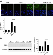
          BV2 microglial cells were pretreated with 2 μg/ml G-Re and stimulated with 1 μg/ml LPS to induce neuroinflammation. To investigate the effect of G-Re on LPS-induced cell signaling, we performed western blotting and immunofluorescence using specific antibodies, such as phospho-p38, COX2, and iNOS.

          Results

          Pretreatment with 2 μg/ml G-Re was neuroprotective against 1 μg/ml LPS-treated microglial cells. The neuroprotective events induced by G-Re treatment in neuroinflammation occurred via the phospho-p38, iNOS, and COX2 signaling pathways in BV2 cells.

          Conclusion

          Taken together, we suggest that G-Re exerts a beneficial effect on neuroinflammatory events in neurodegenerative diseases.

          Related collections

          Most cited references28

          • Record: found
          • Abstract: found
          • Article: not found

          Microglia and inflammation-mediated neurodegeneration: multiple triggers with a common mechanism.

          Inflammation, a common denominator among the diverse list of neurodegenerative diseases, has recently been implicated as a critical mechanism responsible for the progressive nature of neurodegeneration. Microglia are the resident innate immune cells in the central nervous system and produce a barrage of factors (IL-1, TNFalpha, NO, PGE2, superoxide) that are toxic to neurons. Evidence supports that the unregulated activation of microglia in response to environmental toxins, endogenous proteins, and neuronal death results in the production of toxic factors that propagate neuronal injury. In the following review, we discuss the common thread of microglial activation across numerous neurodegenerative diseases, define current perceptions of how microglia are damaging neurons, and explain how the microglial response to neuronal damage results in a self-propelling cycle of neuron death.
            Bookmark
            • Record: found
            • Abstract: found
            • Article: not found

            Inflammatory neurodegeneration and mechanisms of microglial killing of neurons.

            Inflammatory neurodegeneration contributes to a wide variety of brain pathologies. A number of mechanisms by which inflammatory-activated microglia and astrocytes kill neurons have been identified in culture. These include: (1) acute activation of the phagocyte NADPH oxidase (PHOX) found in microglia, (2) expression of the inducible nitric oxide synthase (iNOS) in glia, and (3) microglial phagocytosis of neurons. Activation of PHOX (by cytokines, beta-amyloid, prion protein, lipopolysaccharide, ATP, or arachidonate) causes microglial proliferation and inflammatory activation; thus, PHOX is a key regulator of inflammation. However, activation of PHOX alone causes little or no death, but when combined with iNOS expression results in apparent apoptosis via peroxynitrite production. Nitric oxide (NO) from iNOS expression also strongly synergizes with hypoxia to induce neuronal death because NO inhibits cytochrome oxidase in competition with oxygen, resulting in glutamate release and excitotoxicity. Finally, microglial phagocytosis of these stressed neurons may contribute to their loss.
              Bookmark
              • Record: found
              • Abstract: found
              • Article: not found

              Antidiabetic effects of Panax ginseng berry extract and the identification of an effective component.

              We evaluated antihyperglycemic and anti-obese effects of Panax ginseng berry extract and its major constituent, ginsenoside Re, in obese diabetic C57BL/6J ob/ ob mice and their lean littermates. Animals received daily intraperitoneal injections of Panax ginseng berry extract for 12 days. On day 12, 150 mg/kg extract-treated ob/ob mice became normoglycemic (137 +/- 6.7 mg/dl) and had significantly improved glucose tolerance. The overall glucose excursion during the 2-h intraperitoneal glucose tolerance test decreased by 46% (P < 0.01) compared with vehicle-treated ob/ob mice. The improvement in blood glucose levels in the extract-treated ob/ ob mice was associated with a significant reduction in serum insulin levels in fed and fasting mice. A hyperinsulinemic-euglycemic clamp study revealed a more than twofold increase in the rate of insulin-stimulated glucose disposal in treated ob/ ob mice (112 +/- 19.1 vs. 52 +/- 11.8 micromol x kg(-1) x min(-1) for the vehicle group, P < 0.01). In addition, the extract-treated ob/ob mice lost a significant amount of weight (from 51.7 +/- 1.9 g on day 0 to 45.7 +/- 1.2 on day 12, P < 0.01 vs. vehicle-treated ob/ob mice), associated with a significant reduction in food intake (P < 0.05) and a very significant increase in energy expenditure (P < 0.01) and body temperature (P < 0.01). Treatment with the extract also significantly reduced plasma cholesterol levels in ob/ob mice. Additional studies demonstrated that ginsenoside Re plays a significant role in antihyperglycemic action. This antidiabetic effect of ginsenoside Re was not associated with body weight changes, suggesting that other constituents in the extract have distinct pharmacological mechanisms on energy metabolism.
                Bookmark

                Author and article information

                Journal
                BMC Complement Altern Med
                BMC Complement Altern Med
                BMC Complementary and Alternative Medicine
                BioMed Central
                1472-6882
                2012
                26 October 2012
                : 12
                : 196
                Affiliations
                [1 ]Department of Medical Research, Korea Institute of Oriental Medicine, 483 Expo-ro, Yuseong-gu, Daejeon, 305-811, Republic of Korea
                Article
                1472-6882-12-196
                10.1186/1472-6882-12-196
                3517379
                23102375
                4f4fcf70-ab5c-4269-bb9a-91b691cb9c21
                Copyright ©2012 Lee et al.; licensee BioMed Central Ltd.

                This is an Open Access article distributed under the terms of the Creative Commons Attribution License ( http://creativecommons.org/licenses/by/2.0), which permits unrestricted use, distribution, and reproduction in any medium, provided the original work is properly cited.

                History
                : 16 July 2012
                : 22 October 2012
                Categories
                Research Article

                Complementary & Alternative medicine
                phospho-p38mapk,neuroinflammation,ginsenoside-re (g-re),bv2 microglial cell

                Comments

                Comment on this article