8
views
0
recommends
+1 Recommend
0 collections
    0
    shares
      • Record: found
      • Abstract: found
      • Article: found
      Is Open Access

      Serum IgG-induced microglial activation enhances neuronal cytolysis via the NO/sGC/PKG pathway in children with opsoclonus-myoclonus syndrome and neuroblastoma

      research-article

      Read this article at

      Bookmark
          There is no author summary for this article yet. Authors can add summaries to their articles on ScienceOpen to make them more accessible to a non-specialist audience.

          Abstract

          Background

          Opsoclonus-myoclonus syndrome (OMS) is a rare neurological disease. Some children with OMS also have neuroblastoma (NB). We and others have previously documented that serum IgG from children with OMS and NB induces neuronal cytolysis and activates several signaling pathways. However, the mechanisms underlying OMS remain unclear. Here, we investigated whether nitric oxide (NO) from activated microglias and its cascade contribute to neuronal cytolysis in pediatric OMS.

          Methods

          The activation of cultured cerebral cortical and cerebellar microglias incubated with sera or IgG isolated from sera of children with OMS and NB was measured by the expression of the activation marker, cytokines, and NO. Neuronal cytolysis was determined after exposing to IgG-treated microglia-conditioned media. Using inhibitors and activators, the effects of NO synthesis and its intracellular cascade, namely soluble guanylyl cyclase (sGC) and protein kinase G (PKG), on neuronal cytolysis were evaluated.

          Results

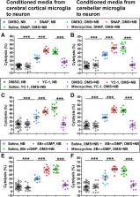
          Incubation with sera or IgG from children with OMS and NB increased the activation of cerebral cortical and cerebellar microglias, but not the activation of astrocytes or the cytolysis of glial cells. Moreover, the cytolysis of neurons was elevated by conditioned media from microglias incubated with IgG from children with OMS and NB. Furthermore, the expression of NO, sGC, and PKG was increased. Neuronal cytolysis was relieved by the inhibitors of NO signaling, while neuronal cytolysis was exacerbated by the activators of NO signaling but not proinflammatory cytokines. The cytolysis of neurons was suppressed by pretreatment with the microglial inhibitor minocycline, a clinically tested drug. Finally, increased microglial activation did not depend on the Fab fragment of serum IgG.

          Conclusions

          Serum IgG from children with OMS and NB potentiates microglial activation, which induces neuronal cytolysis through the NO/sGC/PKG pathway, suggesting an applicability of microglial inhibitor as a therapeutic candidate.

          Related collections

          Most cited references52

          • Record: found
          • Abstract: not found
          • Article: not found

          Microglia-mediated neuroinflammation in neurodegenerative diseases

            Bookmark
            • Record: found
            • Abstract: found
            • Article: not found

            Apolipoprotein E promotes astrocyte colocalization and degradation of deposited amyloid-beta peptides.

            We have previously shown that apolipoprotein E (Apoe) promotes the formation of amyloid in brain and that astrocyte-specific expression of APOE markedly affects the deposition of amyloid-beta peptides (Abeta) in a mouse model of Alzheimer disease. Given the capacity of astrocytes to degrade Abeta, we investigated the potential role of Apoe in this astrocyte-mediated degradation. In contrast to cultured adult wild-type mouse astrocytes, adult Apoe(-/-) astrocytes do not degrade Abeta present in Abeta plaque-bearing brain sections in vitro. Coincubation with antibodies to either Apoe or Abeta, or with RAP, an antagonist of the low-density lipoprotein receptor family, effectively blocks Abeta degradation by astrocytes. Phase-contrast and confocal microscopy show that Apoe(-/-) astrocytes do not respond to or internalize Abeta deposits to the same extent as do wild-type astrocytes. Thus, Apoe seems to be important in the degradation and clearance of deposited Abeta species by astrocytes, a process that may be impaired in Alzheimer disease.
              Bookmark
              • Record: found
              • Abstract: found
              • Article: not found

              Neuroprotection for ischaemic stroke: translation from the bench to the bedside.

              Neuroprotection seeks to restrict injury to the brain parenchyma following an ischaemic insult by preventing salvageable neurons from dying. The concept of neuroprotection has shown promise in experimental studies, but has failed to translate into clinical success. Many reasons exist for this including the heterogeneity of human stroke and the lack of methodological agreement between preclinical and clinical studies. Even with the proposed Stroke Therapy Academic Industry Roundtable criteria for preclinical development of neuroprotective agents for stroke, we have still seen limited success in the clinic, an example being NXY-059, which fulfilled nearly all the Stroke Therapy Academic Industry Roundtable criteria. There are currently a number of ongoing trials for neuroprotective strategies including hypothermia and albumin, but the outcome of these approaches remains to be seen. Combination therapies with thrombolysis also need to be fully investigated, as restoration of oxygen and glucose will always be the best therapy to protect against cell death from stroke. There are also a number of promising neuroprotectants in preclinical development including haematopoietic growth factors, and inhibitors of the nicotinamide adenine dinucleotide phosphate oxidases, a source of free radical production which is a key step in the pathophysiology of acute ischaemic stroke. For these neuroprotectants to succeed, essential quality standards need to be adhered to; however, these must remain realistic as the evidence that standardization of procedures improves translational success remains absent for stroke. © 2012 The Authors. International Journal of Stroke © 2012 World Stroke Organization.
                Bookmark

                Author and article information

                Contributors
                dingxu@bjmu.edu.cn
                wanghuanmin2018@126.com
                Journal
                J Neuroinflammation
                J Neuroinflammation
                Journal of Neuroinflammation
                BioMed Central (London )
                1742-2094
                16 June 2020
                16 June 2020
                2020
                : 17
                : 190
                Affiliations
                [1 ]GRID grid.24696.3f, ISNI 0000 0004 0369 153X, Laboratory of Nutrition and Development, Beijing Pediatric Research Institute, Beijing Children’s Hospital, Capital Medical University, , National Center for Children’s Health, ; No. 56 Nan-li-shi Road, Xi-Cheng District, Beijing, 100045 China
                [2 ]GRID grid.24696.3f, ISNI 0000 0004 0369 153X, Department of Surgical Oncology, Beijing Children’s Hospital, Capital Medical University, , National Center for Children’s Health, ; Beijing, 100045 China
                [3 ]GRID grid.24696.3f, ISNI 0000 0004 0369 153X, Department of Neurobiology, School of Basic Medical Sciences, Beijing Institute for Brain Disorders, , Capital Medical University, ; Beijing, 100069 China
                [4 ]GRID grid.24696.3f, ISNI 0000 0004 0369 153X, Department of Neurology, Beijing Children’s Hospital, Capital Medical University, , National Center for Children’s Health, ; Beijing, 100045 China
                Author information
                http://orcid.org/0000-0002-2529-1217
                Article
                1839
                10.1186/s12974-020-01839-9
                7298801
                32546235
                4f30385b-6c91-4781-9e8e-3225dd10e90f
                © The Author(s) 2020

                Open AccessThis article is licensed under a Creative Commons Attribution 4.0 International License, which permits use, sharing, adaptation, distribution and reproduction in any medium or format, as long as you give appropriate credit to the original author(s) and the source, provide a link to the Creative Commons licence, and indicate if changes were made. The images or other third party material in this article are included in the article's Creative Commons licence, unless indicated otherwise in a credit line to the material. If material is not included in the article's Creative Commons licence and your intended use is not permitted by statutory regulation or exceeds the permitted use, you will need to obtain permission directly from the copyright holder. To view a copy of this licence, visit http://creativecommons.org/licenses/by/4.0/. The Creative Commons Public Domain Dedication waiver ( http://creativecommons.org/publicdomain/zero/1.0/) applies to the data made available in this article, unless otherwise stated in a credit line to the data.

                History
                : 31 December 2019
                : 11 May 2020
                Funding
                Funded by: the BeiJing Talents Fund
                Award ID: 2015000021469G204
                Award Recipient :
                Funded by: the Basic Medicine and Clinical Medicine Cooperation Fund of Capital Medical University
                Award ID: 15JL70
                Award ID: 16JL20
                Award Recipient :
                Funded by: the Scientific Research Cultivation Fund of Capital Medical University
                Award ID: PYZ19030
                Award Recipient :
                Categories
                Research
                Custom metadata
                © The Author(s) 2020

                Neurosciences
                opsoclonus-myoclonus syndrome,neuroblastoma,children,microglial activation,nitric oxide (no),soluble guanylyl cyclase (sgc),protein kinase g (pkg)

                Comments

                Comment on this article