0
views
0
recommends
+1 Recommend
0 collections
    0
    shares
      • Record: found
      • Abstract: found
      • Article: found
      Is Open Access

      Retinal blood flow association with age and weight in infants at risk for retinopathy of prematurity

      research-article

      Read this article at

      Bookmark
          There is no author summary for this article yet. Authors can add summaries to their articles on ScienceOpen to make them more accessible to a non-specialist audience.

          Abstract

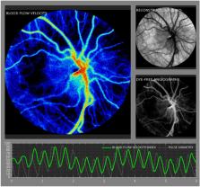
          This prospective study evaluated the relationship between laser speckle contrast imaging (LSCI) ocular blood flow velocity (BFV) and five birth parameters: gestational age (GA), postmenstrual age (PMA) and chronological age (CA) at the time of measurement, birth weight (BW), and current weight (CW) in preterm neonates at risk for retinopathy of prematurity (ROP). 38 Neonates with BW < 2 kg, GA < 32 weeks, and PMA between 27 and 47 weeks underwent 91 LSCI sessions. Correlation tests and regression analysis were performed to quantify relationships between birth parameters and ocular BFV. Mean ocular BFV index in this cohort was 8.8 +/− 4.0 IU. BFV positively correlated with PMA (r = 0.3, p = 0.01), CA (r = 0.3, p = 0.005), and CW (r = 0.3, p = 0.02). BFV did not correlate with GA nor BW (r = − 0.2 and r = − 0.05, p > 0.05). Regression analysis with mixed models demonstrated that BFV increased by 1.2 for every kilogram of CW, by 0.34 for every week of CA, and by 0.36 for every week of PMA ( p = 0.03, 0.004, 0.007, respectively). Our findings indicate that increased age and weight are associated with increased ocular BFV measured using LSCI in premature infants. Future studies investigating the associations between ocular BFV and ROP clinical severity must control for age and/or weight of the infant.

          Related collections

          Most cited references36

          • Record: found
          • Abstract: found
          • Article: not found

          Retinopathy of prematurity.

          The immature retinas of preterm neonates are susceptible to insults that disrupt neurovascular growth, leading to retinopathy of prematurity. Suppression of growth factors due to hyperoxia and loss of the maternal-fetal interaction result in an arrest of retinal vascularisation (phase 1). Subsequently, the increasingly metabolically active, yet poorly vascularised, retina becomes hypoxic, stimulating growth factor-induced vasoproliferation (phase 2), which can cause retinal detachment. In very premature infants, controlled oxygen administration reduces but does not eliminate retinopathy of prematurity. Identification and control of factors that contribute to development of retinopathy of prematurity is essential to prevent progression to severe sight-threatening disease and to limit comorbidities with which the disease shares modifiable risk factors. Strategies to prevent retinopathy of prematurity will depend on optimisation of oxygen saturation, nutrition, and normalisation of concentrations of essential factors such as insulin-like growth factor 1 and ω-3 polyunsaturated fatty acids, as well as curbing of the effects of infection and inflammation to promote normal growth and limit suppression of neurovascular development. Copyright © 2013 Elsevier Ltd. All rights reserved.
            Bookmark
            • Record: found
            • Abstract: found
            • Article: not found

            Screening Examination of Premature Infants for Retinopathy of Prematurity

            This policy statement revises a previous statement on screening of preterm infants for retinopathy of prematurity (ROP) that was published in 2013. ROP is a pathologic process that occurs in immature retinal tissue and can progress to a tractional retinal detachment, which may then result in visual loss or blindness. For more than 3 decades, treatment of severe ROP that markedly decreases the incidence of this poor visual outcome has been available. However, severe, treatment-requiring ROP must be diagnosed in a timely fashion to be treated effectively. The sequential nature of ROP requires that infants who are at-risk and preterm be examined at proper times and intervals to detect the changes of ROP before they become destructive. This statement presents the attributes of an effective program to detect and treat ROP, including the timing of initial and follow-up examinations.
              Bookmark
              • Record: found
              • Abstract: found
              • Article: not found

              Retinopathy of prematurity.

              Retinopathy of prematurity (ROP) is a common blinding disease in children in the developed world despite current treatment, and is becoming increasingly prevalent in the developing world. ROP progresses in two phases. The first phase begins with delayed retinal vascular growth after birth and partial regression of existing vessels, followed by a second phase of hypoxia-induced pathological vessel growth. Two major risk factors of ROP are the use of oxygen and a decreased gestation period. Excessive oxygen contributes to ROP through regulation of vascular endothelial growth factor (VEGF). Suppression of VEGF by oxygen in phase I of ROP inhibits normal vessel growth, whereas elevated levels of VEGF induced by hypoxia in phase II of ROP precipitate pathological vessel proliferation. Insulin-like growth factor 1 (IGF-1) is a critical non-oxygen-regulated factor in ROP. We have found that serum levels of IGF-1 in premature babies directly correlate with the severity of clinical ROP. IGF-1 acts indirectly as a permissive factor by allowing maximal VEGF stimulation of vessel growth. Lack of IGF-1 in preterm infants prevents normal retinal vascular growth in phase I of ROP, despite the presence of VEGF. As infants mature, rising levels of IGF-1 in phase II of ROP allows VEGF stimulated pathological neovascularization. These findings suggest that restoration of IGF-1 to normal levels might be useful in preventing ROP in preterm infants.
                Bookmark

                Author and article information

                Contributors
                jalexander@som.umaryland.edu
                Journal
                Sci Rep
                Sci Rep
                Scientific Reports
                Nature Publishing Group UK (London )
                2045-2322
                4 June 2024
                4 June 2024
                2024
                : 14
                : 12790
                Affiliations
                [1 ]GRID grid.411024.2, ISNI 0000 0001 2175 4264, University of Maryland School of Medicine, ; Baltimore, MD USA
                [2 ]GRID grid.411024.2, ISNI 0000 0001 2175 4264, Department of Ophthalmology and Visual Sciences, , University of Maryland School of Medicine, ; Baltimore, MD USA
                [3 ]GRID grid.411024.2, ISNI 0000 0001 2175 4264, Division of Neonatology, Department of Pediatrics, , University of Maryland School of Medicine, ; Baltimore, MD USA
                [4 ]Department of Biostatistics and Epidemiology, University of Maryland, ( https://ror.org/04rq5mt64) Baltimore, MD USA
                Article
                63534
                10.1038/s41598-024-63534-6
                11150459
                38834830
                4ed58b14-9895-4771-ba63-3f89d2859754
                © The Author(s) 2024

                Open Access This article is licensed under a Creative Commons Attribution 4.0 International License, which permits use, sharing, adaptation, distribution and reproduction in any medium or format, as long as you give appropriate credit to the original author(s) and the source, provide a link to the Creative Commons licence, and indicate if changes were made. The images or other third party material in this article are included in the article's Creative Commons licence, unless indicated otherwise in a credit line to the material. If material is not included in the article's Creative Commons licence and your intended use is not permitted by statutory regulation or exceeds the permitted use, you will need to obtain permission directly from the copyright holder. To view a copy of this licence, visit http://creativecommons.org/licenses/by/4.0/.

                History
                : 29 January 2024
                : 29 May 2024
                Funding
                Funded by: the University of Maryland School of Medicine’s Proposed Research Initiated by Students and Mentors (PRISM) Program
                Funded by: Maryland Industrial Partnerships (MIPS) Program
                Award ID: Grant 7103
                Award Recipient :
                Funded by: National Eye Institute (NEI) of the National Institutes of Health
                Award ID: K23EY03252
                Award Recipient :
                Funded by: Small Business Innovation Research (SBIR)
                Award ID: R43EY030798
                Award Recipient :
                Categories
                Article
                Custom metadata
                © Springer Nature Limited 2024

                Uncategorized
                medical research,paediatric research
                Uncategorized
                medical research, paediatric research

                Comments

                Comment on this article