18
views
0
recommends
+1 Recommend
0 collections
    0
    shares
      • Record: found
      • Abstract: found
      • Article: found
      Is Open Access

      Early stress-induced impaired microglial pruning of excitatory synapses on immature CRH-expressing neurons provokes aberrant adult stress responses

      research-article

      Read this article at

      Bookmark
          There is no author summary for this article yet. Authors can add summaries to their articles on ScienceOpen to make them more accessible to a non-specialist audience.

          SUMMARY

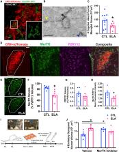
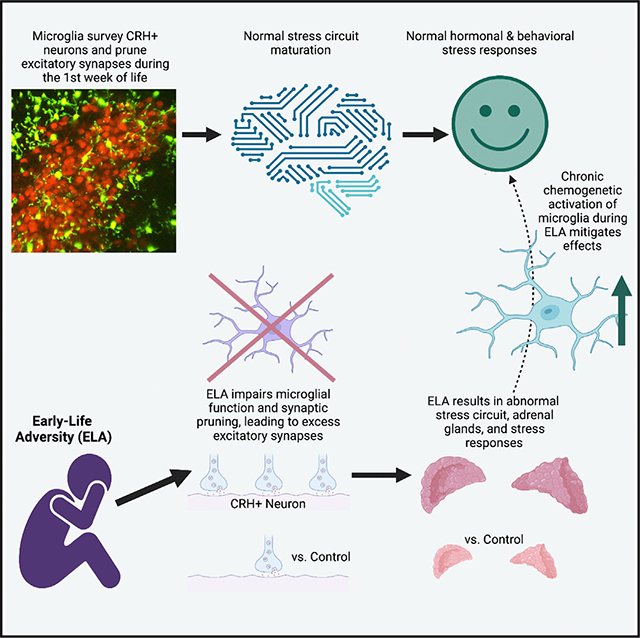
          Several mental illnesses, characterized by aberrant stress reactivity, often arise after early-life adversity (ELA). However, it is unclear how ELA affects stress-related brain circuit maturation, provoking these enduring vulnerabilities. We find that ELA increases functional excitatory synapses onto stress-sensitive hypothalamic corticotropin-releasing hormone (CRH)-expressing neurons, resulting from disrupted developmental synapse pruning by adjacent microglia. Microglial process dynamics and synaptic element engulfment were attenuated in ELA mice, associated with deficient signaling of the microglial phagocytic receptor MerTK. Accordingly, selective chronic chemogenetic activation of ELA microglia increased microglial process dynamics and reduced excitatory synapse density to control levels. Notably, selective early-life activation of ELA microglia normalized adult acute and chronic stress responses, including stress-induced hormone secretion and behavioral threat responses, as well as chronic adrenal hypertrophy of ELA mice. Thus, microglial actions during development are powerful contributors to mechanisms by which ELA sculpts the connectivity of stress-regulating neurons, promoting vulnerability to stress and stress-related mental illnesses.

          In brief

          Early-life adversity (ELA) promotes lifelong aberrant stress responses and vulnerability to mental illnesses. Bolton et al. identify poor dynamics and hypothalamic CRH neurons’ excitatory synapse pruning of ELA microglia, implicating microglial MerTK. Chronic chemogenetic activation of ELA microglia normalized process dynamics, synapse density, and adult hormonal and behavioral stress responses.

          Graphical Abstract

          Related collections

          Most cited references112

          • Record: found
          • Abstract: found
          • Article: not found

          Fiji: an open-source platform for biological-image analysis.

          Fiji is a distribution of the popular open-source software ImageJ focused on biological-image analysis. Fiji uses modern software engineering practices to combine powerful software libraries with a broad range of scripting languages to enable rapid prototyping of image-processing algorithms. Fiji facilitates the transformation of new algorithms into ImageJ plugins that can be shared with end users through an integrated update system. We propose Fiji as a platform for productive collaboration between computer science and biology research communities.
            Bookmark
            • Record: found
            • Abstract: found
            • Article: found
            Is Open Access

            SciPy 1.0: fundamental algorithms for scientific computing in Python

            SciPy is an open-source scientific computing library for the Python programming language. Since its initial release in 2001, SciPy has become a de facto standard for leveraging scientific algorithms in Python, with over 600 unique code contributors, thousands of dependent packages, over 100,000 dependent repositories and millions of downloads per year. In this work, we provide an overview of the capabilities and development practices of SciPy 1.0 and highlight some recent technical developments.
              Bookmark
              • Record: found
              • Abstract: found
              • Article: not found

              An RNA-sequencing transcriptome and splicing database of glia, neurons, and vascular cells of the cerebral cortex.

              The major cell classes of the brain differ in their developmental processes, metabolism, signaling, and function. To better understand the functions and interactions of the cell types that comprise these classes, we acutely purified representative populations of neurons, astrocytes, oligodendrocyte precursor cells, newly formed oligodendrocytes, myelinating oligodendrocytes, microglia, endothelial cells, and pericytes from mouse cerebral cortex. We generated a transcriptome database for these eight cell types by RNA sequencing and used a sensitive algorithm to detect alternative splicing events in each cell type. Bioinformatic analyses identified thousands of new cell type-enriched genes and splicing isoforms that will provide novel markers for cell identification, tools for genetic manipulation, and insights into the biology of the brain. For example, our data provide clues as to how neurons and astrocytes differ in their ability to dynamically regulate glycolytic flux and lactate generation attributable to unique splicing of PKM2, the gene encoding the glycolytic enzyme pyruvate kinase. This dataset will provide a powerful new resource for understanding the development and function of the brain. To ensure the widespread distribution of these datasets, we have created a user-friendly website (http://web.stanford.edu/group/barres_lab/brain_rnaseq.html) that provides a platform for analyzing and comparing transciption and alternative splicing profiles for various cell classes in the brain. Copyright © 2014 the authors 0270-6474/14/3411929-19$15.00/0.
                Bookmark

                Author and article information

                Journal
                101573691
                39703
                Cell Rep
                Cell Rep
                Cell reports
                2211-1247
                9 April 2022
                29 March 2022
                18 April 2022
                : 38
                : 13
                : 110600
                Affiliations
                [1 ]Department of Pediatrics, University of California, Irvine, Irvine, CA, USA
                [2 ]Department of Anatomy/Neurobiology, University of California, Irvine, Irvine, CA, USA
                [3 ]Department of Physiology and Biophysics, University of California, Irvine, Irvine, CA, USA
                [5 ]Département de Médecine Moléculaire, Université Laval, Québec City, QC, Canada
                [6 ]Axe Neurosciences, Centre de recherche du CHU de Québec, Québec City, QC, Canada
                [7 ]Division of Neuroscience, Medical Research Institute, Dundee University, Ninewells Hospital and Medical School, Dundee, UK
                [8 ]These authors contributed equally
                [9 ]Present address: Neuroscience Institute, Center for Neuroinflammation and Cardiometabolic Diseases, Georgia State University, Atlanta, GA, USA
                [10 ]Present address: Convelo Therapeutics, Cleveland, OH, USA
                [11 ]Present address: Division of Medical Sciences, University of Victoria, Victoria, BC, Canada
                [12 ]Lead contact
                Author notes

                AUTHOR CONTRIBUTIONS

                J.L.B. and T.Z.B. designed the experiments. J.L.B., M.S., S.O., S.M.L., A.K.S., C.L.K., B.G.G., and J.C.S. performed the experiments. J.B. and X.B. developed the analytical tools. J.L.B., M.S., S.O., S.M.L., A.K.S., J.B., X.B., C.L.K., B.G.G., and T.Z.B. analyzed the data. J.J.L., D.B., M.-E.T., and M.D.C. provided conceptual advice and access to critical equipment and also supervised collaborative work in their respective laboratories. J.L.B. and T.Z.B. wrote and edited the paper.

                [* ]Correspondence: jbolton@ 123456gsu.edu (J.L.B.), tallie@ 123456uci.edu (T.Z.B.)
                Article
                NIHMS1793839
                10.1016/j.celrep.2022.110600
                9014810
                35354026
                4e53c43f-fe37-4d6b-9958-4a8d1dc2dd32

                This is an open access article under the CC BY-NC-ND license ( http://creativecommons.org/licenses/by-nc-nd/4.0/).

                History
                Categories
                Article

                Cell biology
                Cell biology

                Comments

                Comment on this article